
Introduction
Try to explain how you ride a bicycle. Not the summary version. The actual instructions: the exact lean angle for a right turn at 12 miles an hour, the countersteer your wrists apply when the frame tips left, the dozens of micro-corrections your trunk muscles make every second. You cannot do it. Nobody can. And yet you can ride, flawlessly, after fifteen years off a bike, while holding a conversation about something else entirely.
That gap is the signature of procedural memory. The relationship between the cerebellum and procedural memory is what closes it, and the structure doing most of the quiet work sits at the back of your skull, tucked under the cerebral hemispheres, holding roughly 69 billion neurons in a lump the size of a fist [1]. The cerebellum. Latin for "little brain." A name that has aged badly, because "little" turns out to describe only its volume.
For most of the twentieth century, textbooks filed the cerebellum under motor coordination and moved on. Balance. Posture. Smooth reaching. Useful, but not interesting. Then a mathematician wrote a paper in 1969 predicting exactly how it should learn, a physiologist in Tokyo spent thirteen years proving him half right, and a psychologist in California localized an entire memory to a cluster of cells the size of a pea.
This is the story of how that happened, what the cerebellum actually does when you practise something, and why the field still argues about almost every mechanism involved.
The Structure Everyone Underestimated
Start with the numbers, because they are genuinely strange.
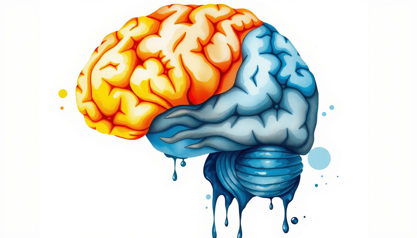
When Suzana Herculano-Houzel and her colleagues dissolved human brains into a uniform soup of cell nuclei and counted them directly, a method called the isotropic fractionator, they arrived at a total of about 86 billion neurons [2]. The cerebral cortex, the wrinkled outer sheet responsible for thought and perception, held about 16 billion of them. The cerebellum held about 69 billion [1].
Read that again. The structure your neuroscience textbook gave four pages contains roughly four out of every five neurons in your head, packed into around a tenth of the brain's mass.
Almost all of them are one cell type: the granule cell. These are among the smallest neurons in the body, with cell bodies five to eight micrometres across, and they are the most numerous neuron on Earth by a wide margin [3]. The same counting work also quietly killed two textbook myths along the way, the "100 billion neurons" figure and the claim that glial cells outnumber neurons ten to one. Neither was ever measured. Both were repeated for decades [4].
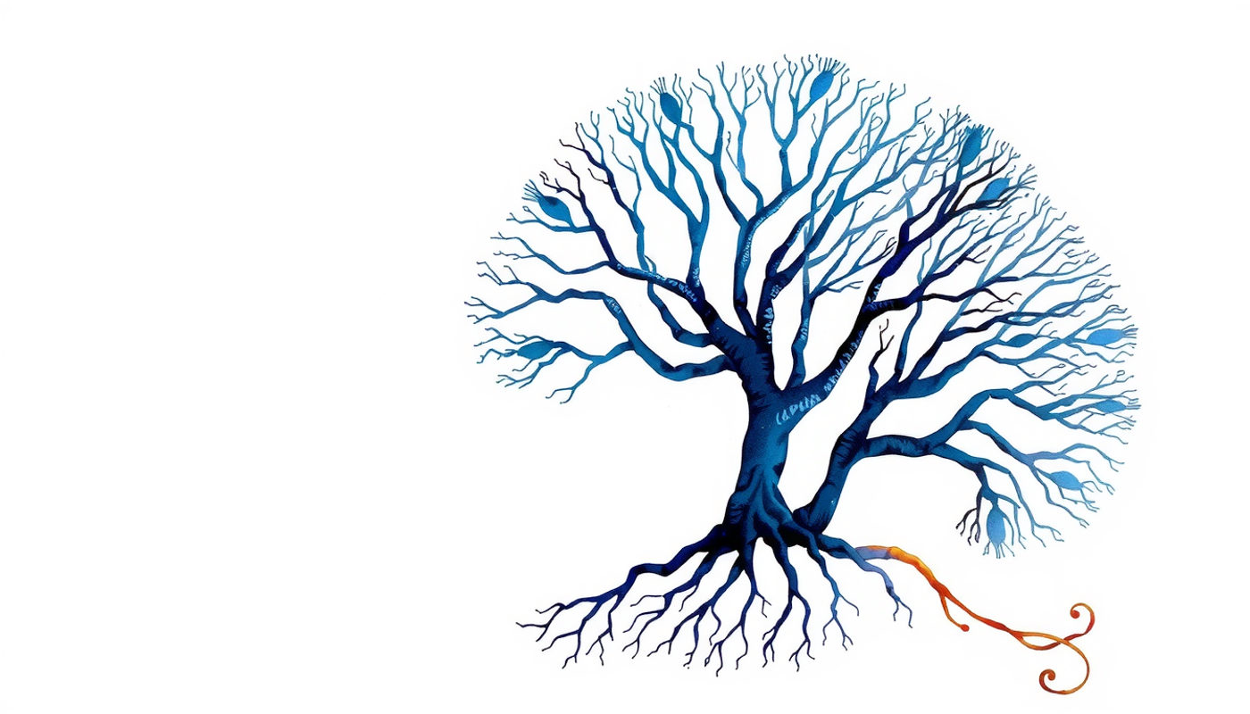
The cerebellar cortex has three layers, and they are almost boringly regular. The bottom layer is packed with granule cells. The middle layer holds a single sheet of Purkinje cells, large neurons with dendritic trees so flat and elaborate they look like pressed ferns. The top layer, the molecular layer, is where granule cell axons run as parallel fibres, crossing the Purkinje dendrites at right angles like telegraph wires strung across a row of trees.
One Purkinje cell receives roughly 175,000 parallel fibre synapses [5]. That figure comes from painstaking electron microscopy work by Napper and Harvey in rat cerebellum, counting synapses one by one.
And it receives exactly one climbing fibre.
That asymmetry, 175,000 inputs of one kind and a single input of another, is the entire puzzle of cerebellar learning in one sentence. It bothered people for years. Then someone worked out what it was for.
From a 1969 Prediction to a 1982 Synapse
David Marr was twenty-four years old and finishing a PhD at Cambridge when he published "A theory of cerebellar cortex" in the Journal of Physiology in 1969 [6]. He had almost no experimental data. He had the wiring diagram, drawn by anatomists over the previous decade, and he had the conviction that a circuit this regular must be computing something specific.
His argument ran roughly like this. Information enters the cerebellum through mossy fibres, which carry signals about body position, movement commands, and sensory context. Those mossy fibres synapse onto the enormous population of granule cells. A few thousand distinct mossy fibre inputs get recoded into patterns spread across billions of granule cells, so that any given situation produces a sparse, nearly unique fingerprint of activity across the parallel fibres.
Marr called these codons. Modern language calls it expansion recoding: take a low-dimensional input, blow it up into a vastly higher-dimensional space, and suddenly patterns that overlapped confusingly become easy to tell apart. It is the same trick used in certain machine learning architectures, arrived at by evolution several hundred million years earlier [7].
Then the climbing fibre. Marr proposed it was a teacher. When a movement goes wrong, the inferior olive, a knot of cells in the brainstem, fires a climbing fibre signal that says, in effect, that was an error. The Purkinje cell that receives it should then change how it responds to whichever parallel fibres happened to be active at that moment.
The theory was elegant, specific, and testable. It was also, in one crucial detail, wrong.
Two years later, an American engineer named James Albus published a strikingly similar model in Mathematical Biosciences [8]. Albus was working on robot control at the time and came at the problem from the direction of pattern classifiers. His circuit was nearly identical to Marr's. His prediction about the synapse was the exact opposite.
Marr thought the climbing fibre should strengthen the active parallel fibre synapses. Albus thought it should weaken them.
Think about which one makes sense. If the climbing fibre signals an error, and the parallel fibres active at that instant helped cause the error, then strengthening them would make the same mistake more likely next time. Weakening them makes it less likely. Albus had the sign right because he understood the signal as a correction rather than a reward.
Nobody could settle it from an armchair. Somebody had to record from the cells.

Masao Ito ran a laboratory in Tokyo and had spent years studying a reflex most people never think about. The vestibulo-ocular reflex, or VOR, keeps your gaze steady when your head moves. Shake your head while reading this sentence and the words stay legible. Your eyes are counter-rotating at the exact speed of your head, automatically, with a delay of about ten milliseconds.
Ito's interest was that the VOR can be recalibrated. Put prism goggles on an animal so the visual world moves the wrong way, and within days the reflex reverses its own gain. That is learning, in a circuit simple enough to trace end to end. And the cerebellar flocculus sits right in the middle of it.
In 1982, Ito's group published the result the field had been waiting thirteen years for. Stimulating parallel fibres and climbing fibres together, repeatedly and in close succession, produced a lasting drop in how strongly that Purkinje cell responded to those parallel fibres [9]. A companion paper the same year showed the depression lasted for at least an hour [10].
They called it long-term depression, or LTD. It is the mirror image of long-term potentiation, the synapse-strengthening process that had already become the leading candidate mechanism for memory in the hippocampus.
Albus was right. Marr's circuit, Albus's sign, Ito's synapse. The field started calling it the Marr-Albus-Ito framework, and for the next three decades it was the standard account of how the cerebellum learns.
What does this mean in practice? Consider what a Purkinje cell is actually doing. It fires constantly, spontaneously, at forty to fifty spikes per second even when nothing is happening. Its output is inhibitory. So a Purkinje cell is a brake, held permanently half-on. Learning, in this system, is not about building new connections. It is about learning precisely when to release the brake.
The climbing fibre, meanwhile, fires at about one spike per second. Roughly one signal for every fifty from the cell's own baseline chatter. It is not carrying information about movement. It is carrying a rare, sharp verdict: that was wrong, adjust.
The Memory in the Pea-Sized Nucleus
While Ito was recording synapses in Tokyo, Richard Thompson in California was chasing something more ambitious. He wanted to find a memory. Not the mechanism of memory in general, but the physical location of one specific learned association, in one specific brain.
He chose a task simple enough to make that possible: eyeblink conditioning. Play a tone. Half a second later, puff air at the eye. The eye blinks at the puff, obviously. Repeat it a few hundred times and something changes. The animal starts blinking at the tone, before the puff arrives, and with the blink timed to peak exactly when the puff is due.
That is a memory. It is measurable to the millisecond, it forms reliably, and it is completely unconscious.
In 1984, Thompson and David McCormick published the finding in Science: lesions of the dentate and interpositus nuclei, the deep cerebellar nuclei buried in the white matter beneath the cerebellar cortex, abolished the conditioned blink completely [11]. The animals still blinked to the air puff. The reflex was fine. They had simply lost the learned response, and could not relearn it.
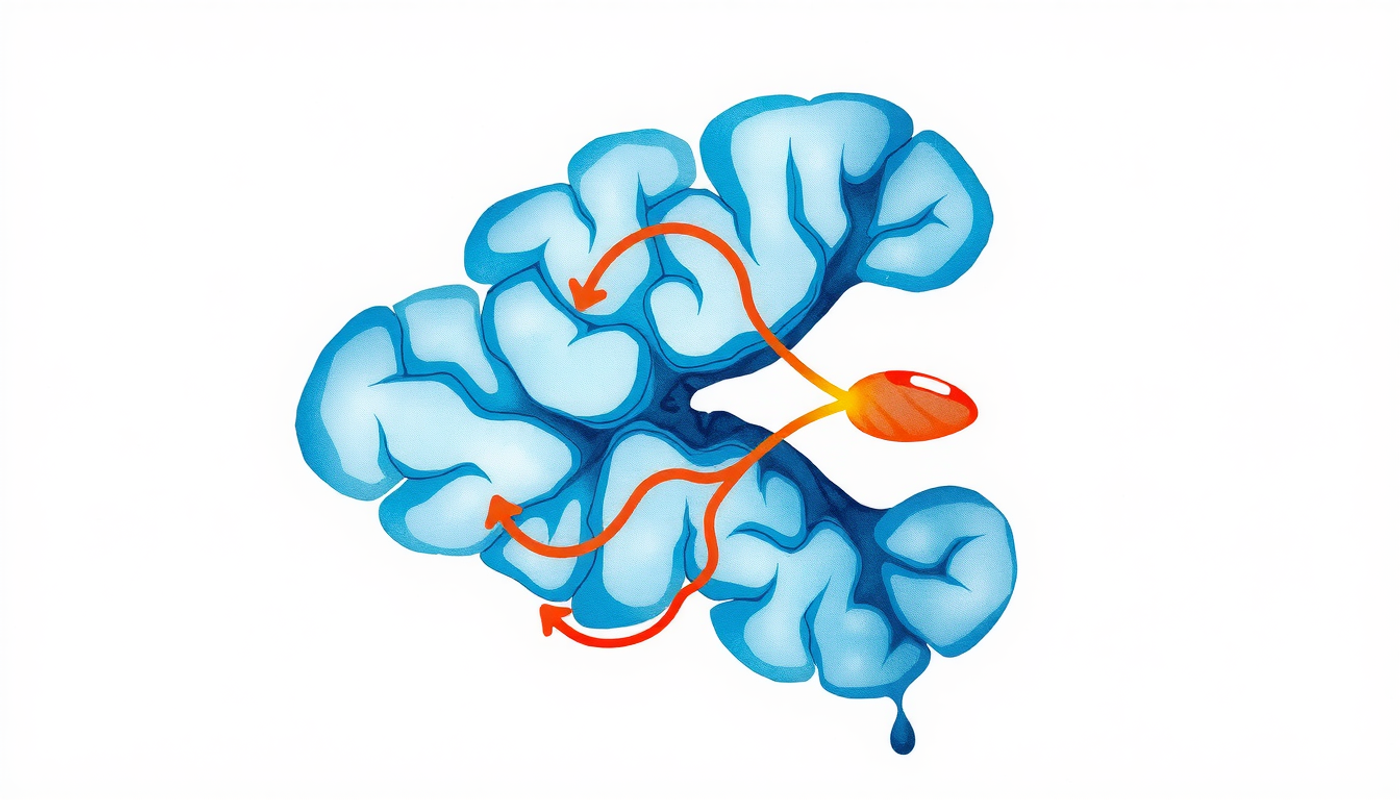
The circuit turned out to be traceable in full. The tone travels via the pontine nuclei in the brainstem and arrives as mossy fibre input. The air puff travels via the trigeminal nerve to the inferior olive and arrives as climbing fibre input. Both converge on the same Purkinje cells and the same corner of the anterior interpositus nucleus. The output leaves through the superior cerebellar peduncle, relays through the red nucleus, and reaches the muscles that close the eyelid [12].
Two streams, one convergence point. The warning signal arrives along one route, the error signal along another, and the Purkinje cell sits where they meet.
Lesions have an obvious weakness as evidence, though. Destroy a region and the animal fails, and you still cannot tell whether you removed the memory or merely the ability to express it.
So in 1993 Thompson's group did something more surgical. During training, they temporarily silenced the interpositus with a drug, then let it recover and tested afterwards [13]. If the memory were forming somewhere upstream and merely passing through the cerebellum on its way out, the animals would show learning once the block wore off. They showed nothing. Then they learned from scratch, at the normal rate, as if the training sessions had never happened.
The trace forms inside the cerebellum. The paper was titled, without hedging, "Localization of a Memory Trace in the Mammalian Brain."
There is a wrinkle in eyeblink conditioning that turned out to matter enormously, and it comes down to a gap of a few hundred milliseconds.
In delay conditioning, the tone stays on until the air puff arrives. The two stimuli overlap. This version is purely cerebellar. Animals with the rest of the forebrain surgically removed still learn it.
In trace conditioning, the tone stops, then there is a silent gap, then the puff. Nothing physically connects them. The brain has to hold a representation of the vanished tone across empty time. And that version requires the hippocampus and the prefrontal cortex on top of the cerebellum.
Robert Clark and Larry Squire tested what this meant in humans, and the result is one of the cleaner demonstrations of what conscious awareness is actually for [14]. They ran both versions on volunteers while distracting them with a silent film, then asked afterwards whether they had noticed any relationship between the tone and the puff. Delay conditioning worked regardless. Participants who had no idea a relationship existed still developed anticipatory blinks. Trace conditioning worked only in people who could report the relationship out loud.
Amnesic patients with damage to the medial temporal lobe, the region containing the hippocampus, showed the same split. Delay conditioning, normal. Trace conditioning, absent.
This is the same dissociation that Henry Molaison made famous. After surgery removed both his medial temporal lobes in 1953, Molaison could not form new conscious memories, yet he learned motor skills at an ordinary rate and improved across days he had no memory of experiencing [15]. Neal Cohen and Larry Squire formalized the principle in 1980 with amnesic patients learning mirror reading, a skill that improved steadily while the patients denied ever having done the task [16].
Knowing how and knowing that are separate systems. And knowing how does not need you to be paying attention.
What does this mean for anyone learning a physical skill? The part of practice that builds automaticity is not the part you are consciously monitoring. You can attend, evaluate, and narrate all you like, and that effort feeds a different memory system than the one gradually smoothing out your timing.
Here is a question that should have a complicated answer. How does the cerebellum know that the puff is coming in 250 milliseconds and not 400?
The conditioned blink is exquisitely timed. It peaks right when the air puff is due. Change the interval during training and the blink shifts to match. The optimal interval sits somewhere around a quarter of a second, and outside a range of roughly 150 to 500 milliseconds the learning degrades badly.
For years the obvious explanation was a network. Some circuit of interconnected neurons passing activity along in a chain, functioning as a clock, with the Purkinje cell reading off the appropriate tick.
Germund Hesslow's group at Lund University demolished that idea in 2014 [17]. They trained ferrets using direct electrical stimulation of parallel fibres as the conditioned signal, bypassing the tone entirely. Then they applied drugs blocking the inhibitory interneurons in the cerebellar cortex, removing the local network. Then they recorded from single Purkinje cells.
The cells still learned. And they still produced adaptively timed responses, a pause in firing that began after the right delay and peaked at the right moment. Individual cells, isolated from their network, had learned to measure a quarter of a second.
The timing mechanism is intrinsic to the cell.
That is a genuinely odd result, and its implications are still being worked out. A single neuron can apparently be trained to hold a specific interval, which suggests something happening inside the cell, in its molecular machinery, rather than in the wiring between cells. Ivry and Keele had argued back in 1989 that the cerebellum functions as a general-purpose timing system, used for motor and perceptual tasks alike [18]. They were arguing about a network. It may be a population of individually competent clocks.
The Theory That Started Failing
For thirty years, cerebellar LTD was the answer. Then the mice arrived.
In 2011, Martijn Schonewille and colleagues in Chris De Zeeuw's laboratory in Rotterdam published a paper in Neuron with a title that reads like a polite demolition: "Reevaluating the Role of LTD in Cerebellar Motor Learning" [19].
They had three separate lines of genetically modified mice in which the molecular machinery for parallel fibre LTD was blocked, each by a different route. If LTD is the substrate of cerebellar learning, these animals should not learn.
They learned. VOR adaptation, normal. Eyeblink conditioning, normal. Locomotion, normal.
The response was immediate and heated. Ito's group argued that LTD could still be induced in those mice using different stimulation protocols, and pointed out that several other LTD-deficient mutant lines do show clear learning deficits. The disagreement has never fully closed.
What emerged instead is messier and probably closer to the truth. Cerebellar learning is not one synapse. Gao, van Beugen and De Zeeuw laid out the alternative in 2012 [20]: plasticity at the parallel fibre synapse in both directions, plasticity at the mossy fibre to granule cell synapse, changes in the intrinsic excitability of Purkinje cells, plasticity in the molecular layer interneurons, and plasticity in the deep cerebellar nuclei. Multiple mechanisms, distributed across the circuit, working together.
De Zeeuw pushed this further in 2021 with a framework that divides the cerebellar cortex into two kinds of microzone [21]. Upbound zones, where Purkinje cells have relatively low baseline firing and learning drives their activity up. Downbound zones, where baseline firing is high and learning drives it down. The two types differ in molecular markers, in physiology, and in which behaviours they support. Same circuit diagram, opposite learning rules, side by side in the same structure.
Jennifer Raymond and Javier Medina summarized the state of play in 2018 with a phrase worth remembering: the cerebellum is a supervised learning machine, but the supervision is distributed across more sites than anyone originally imagined [22].
Fifty years after Marr, a retrospective review by Mitsuo Kawato and colleagues put it plainly [23]. The core architecture Marr described has held up remarkably well. The single-synapse learning rule has not.
Where the Memory Moves After You Sleep on It
One of the strangest findings in cerebellar research is that the memory does not stay put.
Shutoh and colleagues in Nagao's laboratory trained mice on optokinetic reflex adaptation, a close cousin of the VOR, and then went looking for where the learning lived [24]. After about an hour of training, the memory was in the cerebellar cortex. Damage the flocculus and the learning vanished. But after several days of repeated training, damaging the same cortex did nothing. The behaviour survived. The memory had relocated to the vestibular nuclei downstream.
Kassardjian's group found the same pattern in cats using VOR adaptation, and titled the paper accordingly: "The Site of a Motor Memory Shifts with Consolidation" [25].
Then Okamoto and colleagues asked what drives the transfer. They blocked protein synthesis in the cerebellar cortex during the consolidation window, and the memory stayed stuck in the cortex instead of moving [26]. The cortex has to actively build something in order to hand the memory over.
The emerging picture is a division of labour across time. The cerebellar cortex acquires quickly, because it has the climbing fibre error signal and the enormous granule cell population for pattern separation. The deep nuclei store durably, because they sit at the output stage and can hold a stable bias without needing the fine-grained learning machinery.
Cortex learns. Nuclei keep.
This is worth sitting with if you have ever noticed that a physical skill feels differently owned after a few weeks than it did on day one. The early version is fast, flexible, and fragile. The consolidated version is slower to change and much harder to lose. Those are not just descriptions of your subjective experience. They may correspond to two anatomically distinct storage sites.
The last entry on that list is the one that changes the shape of everything above it.
The 2024 Result That Reframed the Question
For decades, studies of cerebellar patients performing motor adaptation tasks produced a confusing spread of results. Some found devastating impairments. Some found mild ones. Everyone was using broadly similar paradigms, usually force-field adaptation, where a robotic arm pushes sideways against your reaching movement and you gradually learn to counter it.
Maurice Smith and Reza Shadmehr reported in 2005 that patients with cerebellar degeneration were severely impaired at this, while patients with Huntington's disease, a basal ganglia disorder, learned normally [27]. Clean dissociation. But other groups running the same task found deficits of only 30 to 40 percent.
Alkis Hadjiosif, Tricia Gibo and Maurice Smith went back to the raw data from several of those studies and noticed something nobody had thought to check: the spacing between trials.
Their 2024 paper in PNAS carries a title that sounds almost too neat [28]. "The cerebellum acts as the analog to the medial temporal lobe for sensorimotor memory."
Here is the argument. Sensorimotor memory has two components. One decays fast, with a time constant of 15 to 20 seconds, and can never lead to long-term retention. The other is temporally persistent, stable for a minute or more, and is what actually accumulates into durable learning.
Across the pooled patient data, the overall learning deficit in severe cerebellar degeneration was a modest 35 percent. But when they separated trials by spacing, the picture split apart. On trials following short intervals of five to eight seconds, the deficit was about 34 percent. On trials following long breaks of 25 seconds or more, the deficit was 86 percent.
Cerebellar patients were not failing to learn. They were failing to keep what they learned past the point where the volatile component decayed away.
That maps precisely onto what the medial temporal lobe does for facts and events. Damage the hippocampus and short-term memory works fine while long-term memory formation collapses. Damage the cerebellum and short-term sensorimotor memory works fine while long-term sensorimotor memory collapses.
Two gateways. Two categories of memory. Same architectural principle.
The authors are careful about the limits of their own claim. They note explicitly that not every form of non-declarative learning depends on the cerebellum. This is a statement about sensorimotor memory, not about procedural memory as a whole.
Still. It explains fifteen years of contradictory literature with one variable that nobody was controlling.
The Other Half of the System
The cerebellum is not doing this alone. Procedural memory is a partnership, and the other partner is the basal ganglia, a set of deep structures including the striatum that sit near the centre of the brain.
The classical division of labour goes like this. The cerebellum runs supervised learning, driven by error signals about the difference between what was predicted and what happened. The basal ganglia run reinforcement learning, driven by dopamine signals about the difference between expected and received reward.
Julien Doyon and colleagues mapped this onto motor learning with a specific prediction [29]. In the early stages of learning, both systems engage together along with cortex. But as learning consolidates, they separate by task type. Motor sequence learning, the kind involved in typing a passcode or playing a scale, shifts toward cortico-striatal circuits. Motor adaptation, the kind involved in adjusting to a new racket weight or a shifted visual field, shifts toward cortico-cerebellar circuits [30].
That framework has been useful for twenty years. It is also getting harder to defend in its clean form.
Andreea Bostan and Peter Strick published anatomical work in 2018 that should have caused more of a stir than it did [31]. Using trans-neuronal virus tracing, which follows connections across synapses, they showed that the subthalamic nucleus of the basal ganglia projects to the cerebellar cortex, and the dentate nucleus of the cerebellum projects to the striatum. Two synapses in each direction. The systems that textbooks drew as parallel and independent are directly wired together.
And then the cerebellum turned out to care about reward.
Mark Wagner and colleagues in Liqun Luo's laboratory used two-photon calcium imaging, a technique that lets you watch hundreds of individual neurons light up in a living animal, to record granule cells in mice performing a reaching task for a sugar reward [3]. Some granule cells responded to reward. Others responded specifically to reward being omitted. Others fired in anticipation of reward. None of it tracked the licking movements. These were not motor signals.
Two years later, Ilaria Carta and colleagues found a direct excitatory projection from the cerebellar nuclei to the ventral tegmental area, the dopamine hub at the centre of the brain's reward system [32]. Optogenetically activating it was rewarding in itself. Mice spent most of their time in whichever corner of the box triggered the stimulation. Silencing it disrupted normal social preference.
A structure supposedly dedicated to motor error correction has a private line to the dopamine system. Dimitar Kostadinov and Michael Häusser reviewed the accumulating evidence in 2022 and concluded that reward signals in the cerebellum are real, widespread, and not yet explained by any existing theory [33].
The table above is a useful simplification. Treat it as scaffolding rather than settled fact.
What Cerebellar Patients Can Still Do
The clinical literature is where the theory meets people, and it is more nuanced than the lesion studies suggest.
Patients with cerebellar degeneration, most often from spinocerebellar ataxia, a group of inherited disorders that progressively kill Purkinje cells, do show clear adaptation deficits. Ya-Weng Tseng and colleagues demonstrated that these patients specifically fail to use sensory prediction errors, the mismatch between where the arm was expected to go and where it went [34]. Rabe and colleagues found that visuomotor rotation deficits and force-field deficits correlate with damage in different cerebellar zones, suggesting the structure is not uniformly responsible for everything [35].
Susanne Morton and Amy Bastian tested walking, using a split-belt treadmill where the two legs move at different speeds [36]. Healthy people gradually build a new gait pattern that anticipates the mismatch. Cerebellar patients could not build that predictive adjustment. But their reactive, within-step corrections were intact. They could respond. They could not predict.
That distinction runs through the whole clinical picture. The cerebellum is a prediction machine, and what damage removes is the anticipation, not the reaction.
Now for the part that gets less attention. Cerebellar patients retain a lot.
Sarah Criscimagna-Hemminger and colleagues found that when a perturbation is introduced gradually rather than abruptly, patients adapt much better [37]. Small errors are handled more successfully than large ones.
Amy Bastian's group also showed that cerebellar patients can learn from binary reinforcement, simple hit-or-miss feedback with no information about direction [38]. They learn less efficiently, and the authors attributed most of that inefficiency to excess motor noise. Their movements are so variable that the exploration process becomes unreliable. Chris Miall and Joseph Galea offered a commentary arguing the interpretation may be too generous to the noise account [39]. A follow-up study added matched artificial noise to healthy participants and found it slowed learning, but not nearly to patient levels [40]. Something beyond noise is going on.
And patients can use explicit strategy. Jordan Taylor, John Krakauer and Richard Ivry demonstrated in 2014 that what looks like a single adaptation process is actually two [41]. There is a deliberate, conscious re-aiming component, and there is an implicit, unconscious recalibration component. They can be separated experimentally by asking participants where they intend to aim. The implicit component is stubbornly rigid. It reaches roughly the same asymptote regardless of how large the error is. It is also the component that depends on the cerebellum.
What does this mean for anyone recovering from a cerebellar injury, or coaching someone who is? Gradual change beats abrupt change. Explicit instruction still works. And the deficit is specific enough that it should be trained around rather than trained through.
The Sleep Story Nobody Should Repeat Uncritically
Somewhere along the way, one number escaped the laboratory and became folklore.
In 2002, Matthew Walker, Robert Stickgold and colleagues published a study in Neuron using the motor sequence task, in which participants type a five-digit sequence as fast and accurately as possible [42]. Train in the morning, test twelve hours later while awake: no improvement. Train in the evening, sleep, test in the morning: about 20 percent faster with no loss of accuracy.
Sleep, apparently, improved a motor skill with no additional practice. The effect correlated with stage 2 non-REM sleep. A later nap study found the gain did not track spindle activity over the learning hemisphere on its own. It tracked the difference between the two hemispheres, a relationship that disappeared when either side was measured alone [43].
It is a beautiful result. It is also, after two decades of argument, seriously contested.
Timothy Rickard and colleagues pointed out a problem with how the task was scored [44]. Performance was averaged across long training blocks. Within any long block, people slow down as they fatigue, a phenomenon called reactive inhibition. So the end-of-training average is depressed by fatigue, and the next-morning average, taken when the participant is fresh, looks higher by comparison. When Rickard's group ran the study with short blocks that minimise fatigue, the overnight gain disappeared.
Their paper was titled "Sleep does not enhance motor sequence learning."
A meta-analysis by Steven Pan and Rickard in 2015 looked across the literature and found little reliable evidence for a sleep-specific enhancement once these confounds were controlled [45]. The balance of opinion has shifted substantially toward scepticism.
None of this means sleep is irrelevant to memory consolidation. It means the specific claim about a 20 percent overnight motor speed boost should be treated as disputed rather than established. Andrew Jackson and Wei Xu reviewed cerebellar involvement in sleep-dependent memory in 2023 and found real evidence of cerebellar activity and replay during sleep [46]. What that activity accomplishes behaviourally is still open.
Note also which kind of learning is at stake. The contested gains are in sequence learning, which leans on the basal ganglia. Cerebellar adaptation tasks show relatively little sleep-specific offline improvement. The two systems appear to consolidate differently, and lumping them together is part of what created the confusion.
When the Little Brain Turns Out to Think
In 1998, Jeremy Schmahmann and Janet Sherman published a paper in Brain describing twenty patients with damage confined to the cerebellum [47]. What they reported was not a movement disorder.
The patients showed impaired executive function, planning problems, difficulty shifting between tasks. Impaired spatial cognition. Language problems including reduced verbal fluency and abnormal prosody, the melody of speech. And personality change, with flattened affect or disinhibited behaviour.
They named it the cerebellar cognitive affective syndrome. Schmahmann's explanatory idea was that the cerebellum performs one computation, applied everywhere, and that damaging it produces a "dysmetria of thought" in the same way it produces dysmetria of movement. Movements overshoot. Thoughts overshoot too.
The syndrome now has a validated clinical scale, developed by Franziska Hoche and colleagues, which makes it diagnosable rather than merely describable [48].
The theoretical bridge from motor control to cognition runs through internal models. Masao Ito, the same physiologist who found LTD, argued in 2008 that the cerebellum builds predictive models of whatever it is connected to [49]. Connected to the motor cortex, it models limb dynamics. Connected to the prefrontal cortex, it models thought. Same computation, different content.
Arseny Sokolov, Chris Miall and Richard Ivry developed this into a framework of adaptive prediction spanning movement and cognition [50]. Court Hull went further in a 2020 review, arguing that the cerebellum generates and tests predictions about reward and other non-motor variables, not just movement errors [51].
Not everyone is convinced. The sceptical position is that many apparent cognitive deficits in cerebellar patients could reflect timing problems or motor demands hidden inside the cognitive tasks. Speech tasks require articulation. Working memory tasks often require sequencing. It is difficult to design a cognitive test with no temporal or motor component at all.
The honest summary: a cerebellar contribution to cognition is now widely accepted, its magnitude is disputed, and whether it reflects one universal computation or many specialized ones is unresolved.
For procedural memory specifically, this matters because skills are not only motor. Grammar, mathematical procedure, chess pattern recognition, clinical diagnosis. All of these become fast and automatic with practice, all of them show the signature of procedural learning, and all of them involve cerebellar loops with the relevant cortical regions.
Eight Arguments the Field Has Not Settled
Scientific writing about the brain tends to smooth over disagreement. This topic does not deserve that treatment, because the disagreements are where the interesting work is happening.
Is LTD necessary for cerebellar motor learning? The evidence says no, not strictly. LTD-blocked mice learn [19]. But several other LTD-deficient lines show real deficits, and the induction protocols matter. The consensus has moved to distributed plasticity across many sites [20]. What remains open is the relative causal weight of each site.
Where does the memory actually live? Cortex first, then deep nuclei, on a timescale of days [24]. That much is reasonably solid for VOR-type adaptation. Whether it generalises to all cerebellar learning, and what the molecular transfer mechanism is, is not resolved.
Does the cerebellum genuinely contribute to cognition, or are those findings motor confounds? Anatomy, clinical syndrome data, and imaging all support a real contribution [48]. The sceptics have a fair point about task design. Probably real, probably modulatory rather than primary.
Are sleep-dependent motor gains real? Contested, and the sceptical case is strong [45]. Sleep may stabilise rather than enhance.
Do cerebellar patients fail at reinforcement learning because of motor noise or because of a direct cerebellar role in reward? Both, in unknown proportions [40]. The reward findings in mice make the direct-role account harder to dismiss.
Does the cerebellum contribute to sequence learning independently of execution? Unclear. Separating learning from performance in patients with movement disorders is genuinely hard.
How clean is the cerebellum-basal ganglia division of labour? Less clean every year. The disynaptic connections are established [31]. The reward signals are established [3]. The tidy dichotomy is a teaching tool, not a description.
Is procedural memory one system? Almost certainly not. Adaptation, sequence learning, conditioning, and habit formation dissociate from one another neurologically. The umbrella term is behaviourally convenient and neurobiologically misleading.
That last point deserves emphasis. When a textbook says procedural memory depends on the cerebellum and basal ganglia, it is compressing at least four distinguishable learning systems into one phrase.
What Any of This Means for Practice
Strip away the anatomy and a few things follow reasonably directly from the evidence.
Practice needs errors. The cerebellum learns from sensory prediction error, the gap between what was expected and what occurred [34]. Practice that eliminates error, by making the task too easy or providing so much guidance that no mismatch ever occurs, removes the teaching signal. The discomfort of getting it wrong is not a side effect of learning. It is the input.
But error size matters. Very large errors adapt poorly. Gradual perturbations produce better learning than abrupt ones, particularly in impaired systems [37]. Incremental difficulty increases are not just gentler. They are mechanistically better matched to how the system updates.
Timing is a learned parameter, not a by-product. The cerebellum encodes intervals with millisecond precision, and single cells can hold specific durations [17]. Practising a musical passage slowly and then speeding it up is not simply practising the same thing faster. Different intervals are, to some extent, different learned quantities.
Spacing between attempts probably matters more than anyone realised. The 2024 finding that cerebellar contribution shows up specifically at long inter-trial intervals implies that the durable component of sensorimotor memory only becomes visible once the volatile component has decayed [28]. Massed repetition may look better during a session while building less that survives it.
And explicit understanding is a separate channel. Conscious re-aiming and implicit recalibration are distinct processes with different properties [41]. Understanding a technique intellectually will not recalibrate the implicit system. Only repetition with feedback does that. Both are useful. Neither substitutes for the other.
None of these are dramatic prescriptions. They are constraints, derived from a system that has been doing supervised error correction for several hundred million years and has no interest in your opinions about how learning should work.
Conclusion
The cerebellum spent a century filed under motor coordination. It turned out to hold four out of every five neurons in the brain, to implement a learning rule that a graduate student predicted from a wiring diagram in 1969, to store timed memories inside individual cells, to talk directly to the dopamine system, and to serve as a gateway for durable skill in the same way the hippocampus serves as a gateway for durable fact.
The story is not finished, and the unfinished parts are visible in the literature if you look. A theory that dominated for thirty years is now considered incomplete. A famous number about sleep is under sustained attack. A tidy division of labour between two brain systems is dissolving under anatomical evidence. These are not failures. This is what a field looks like when it is still moving.
What holds up is the shape of the thing. Somewhere behind and below the thinking part of your brain sits a structure that watches every movement you make, compares it against what was predicted, and quietly adjusts. It does this without asking permission and without reporting back. It learns from being wrong, which is the only reliable teacher any learning system has ever had.
You cannot describe how you ride a bike because the knowledge was never stored in a form that words can reach. It is written in the timing of a few thousand cells learning to release a brake at exactly the right instant.
That is a strange kind of memory to carry around. It is also, most days, the kind you depend on most.
Frequently Asked Questions
Where is procedural memory stored in the brain?
Procedural memory does not sit in one place. Motor adaptation and timed conditioning depend heavily on the cerebellum and its deep nuclei. Sequence learning and habit formation depend more on the basal ganglia and striatum. Motor cortex handles execution. These systems dissociate from one another in patients with different lesions.
Can you lose procedural memory the way you lose factual memory?
Procedural memory is more resilient. Amnesic patients unable to form new conscious memories still learn motor skills at normal rates and improve across sessions they cannot recall. It is impaired instead by damage to the cerebellum, which disrupts adaptation and prediction, or to the basal ganglia, as in Parkinson's disease.
What is the difference between delay and trace conditioning?
In delay conditioning the warning signal overlaps with the outcome, and learning depends only on the cerebellum. In trace conditioning a silent gap separates them, which additionally requires the hippocampus and prefrontal cortex. In humans, trace conditioning only works if the person consciously notices the relationship. Delay conditioning works without awareness.
How long does it take for a skill to become automatic?
There is no fixed number. It depends on task complexity, practice quality, and how consistently errors are available to learn from. Evidence suggests the memory itself relocates during consolidation, moving from cerebellar cortex to deep nuclei over days of repeated training, which may correspond to the shift from fragile to durable skill.
Does the cerebellum do anything besides movement?
Yes, though the extent is debated. Patients with cerebellar-only damage show executive dysfunction, spatial deficits, language problems, and personality changes, described as the cerebellar cognitive affective syndrome. Cerebellar neurons also encode reward expectation and project directly to the brain's dopamine system, which no purely motor theory predicts.


