
Introduction
A seventy-year-old woman stands in her kitchen. She walked in with a purpose. Now she cannot recall what it was. Thirty seconds pass. Nothing. She laughs it off. But somewhere in the back of her mind, a question stirs: is this normal, or is something wrong?
That question sits at the center of one of neuroscience's most active research areas. How aging changes memory is not a simple story of decline. It is a story of selective loss and surprising preservation. Some memory systems fall apart. Others stay intact for decades. A few actually get better with age [1]. In 2025, the largest brain-imaging study ever conducted on this topic pooled 13 longitudinal datasets, 3,737 healthy adults, and over 10,000 MRI scans. What the researchers found overturned decades of assumptions about which parts of the brain drive memory loss [2].
This article tells the story of how scientists discovered what happens inside the aging brain when memories start to slip. It begins with a woman named Brenda Milner and a patient who could not form new memories. It moves through decades of brain scans, dendritic spines, neurotransmitter losses, and a fierce debate about when decline actually starts. And it ends with something unexpected: evidence that a significant portion of age-related memory loss may be preventable.

The Hippocampus: Where the Story Begins
The modern science of memory and aging traces back to one patient. His initials were H.M. In 1953, a neurosurgeon named William Scoville removed large portions of Henry Molaison's medial temporal lobes, including most of his hippocampus, to treat severe epilepsy. The seizures stopped. But so did something else. H.M. could no longer form new memories [3].
Brenda Milner, a neuropsychologist at the Montreal Neurological Institute, studied H.M. for decades. She found something remarkable. He could remember his childhood. He could hold a conversation. He could learn new motor skills without any awareness of having practiced them. But he could not remember meeting Milner, even after hundreds of sessions. Every visit was the first visit.
H.M. proved that the hippocampus, a seahorse-shaped structure deep in each temporal lobe, is essential for converting short-term experiences into lasting memories. Without it, the brain can still access old memories and learn certain skills. But new conscious memories cannot form.
Why does this matter for aging? Because the hippocampus is one of the brain structures most vulnerable to age-related shrinkage. MRI studies show it loses volume at roughly 1 to 2 percent per year after age 50 [4]. The dentate gyrus, a subregion of the hippocampus responsible for generating new neurons, slows its production rate with each passing decade. Some researchers believe new-neuron production drops to near zero in old age [5]. Others have found evidence that it persists, albeit at lower rates, well into the eighth decade of life [6].
This disagreement is more than academic. If the hippocampus continues producing new neurons throughout life, there may be ways to boost that production. If it does not, the strategies for protecting memory shift entirely.
The Mega-Analysis That Changed Everything
For decades, the working assumption was simple: hippocampus shrinks, memory declines. Fix the hippocampus, fix the problem. But in 2025, a team of researchers from Norway, Sweden, Germany, the UK, and the US published a study that complicated this picture dramatically.
Diego Vidal-Piñeiro, together with Anders Fjell, Kristine Walhovd, and Lars Nyberg, pooled data from 13 longitudinal studies. Their dataset was enormous: 3,737 cognitively healthy adults, 10,343 MRI scans, 13,460 memory tests [2]. Instead of looking at one brain region at a time, they estimated structural change across 166 cortical and subcortical regions simultaneously.
Three findings stood out. First, the relationship between brain shrinkage and memory decline is not linear. It follows a curve. People with average or below-average rates of brain atrophy showed little connection between structural change and memory loss. The coupling only became strong in individuals whose brains were shrinking faster than normal. Second, memory decline is not driven by the hippocampus alone. The hippocampus showed the strongest single association, yes. But widespread cortical and subcortical regions contributed. Memory loss in aging reflects a broad structural vulnerability across the entire brain, not damage in one spot. Third, the brain-memory coupling gets stronger with age. In participants in their fifties, the connection between atrophy and memory was weak. By the eighties, it was moderate to strong.
What about genetics? APOE ε4 carriers, who carry the strongest common genetic risk factor for Alzheimer's disease, did show steeper brain shrinkage and faster memory decline. But the ε4 allele did not change the brain-memory relationship itself. Whether someone carried the risk gene or not, the coupling between atrophy and memory loss followed the same pattern.
The implication is significant. Memory aging is not about one broken part. It is about a system gradually losing coherence across many regions at once.
Not All Memories Age the Same Way
Here is something that rarely makes it into popular articles about aging and memory. "Memory" is not one thing. It is a collection of distinct systems that rely on different brain circuits and age at very different rates.
Episodic memory, the ability to recall specific events bound to a time and place, is the most vulnerable. Remembering what you had for breakfast. Recalling where you parked. Knowing what someone said in a meeting last week. These depend heavily on the hippocampus and prefrontal cortex, and they decline measurably from the fifties onward [7].
Working memory, the mental scratch pad that holds information for a few seconds while you use it, also declines. Not dramatically, but consistently. A young adult can hold about seven items in working memory. By the seventies, that number drops to about five or six.
Source memory, remembering where you learned something or who told you, weakens significantly. This creates a specific problem: older adults may recall a fact correctly but misattribute where they learned it, making them more susceptible to misinformation [8].
But semantic memory, general world knowledge and vocabulary, is remarkably resilient. It actually improves well into the sixties and seventies [9]. A seventy-year-old typically knows more words and more facts than a thirty-year-old. Procedural memory, the ability to ride a bicycle or play a musical instrument, stays largely intact. And implicit memory, the unconscious influence of past experiences on current behavior, shows minimal age-related decline.
The practical meaning? When an older person forgets where they put their keys but can explain in detail how a car engine works, that is not contradictory. Two different memory systems are at play, and only one of them has weakened.
Moshe Naveh-Benjamin at the University of Missouri proposed one of the most influential explanations for why episodic memory suffers most. His Associative Deficit Hypothesis, published in 2000, argues that older adults are specifically impaired at binding the pieces of an experience together [8]. They can remember a face. They can remember a name. But connecting the face to the name, the item to its context, the event to its time? That binding process weakens. In experiments, older adults remembered individual items nearly as well as younger adults but performed significantly worse when asked to recall which items had been paired together [10].

Synapses That Shrink, Not Neurons That Die
For most of the twentieth century, scientists assumed that aging killed brain cells. Fewer neurons meant fewer memories. It was a neat, intuitive story. It was also wrong.
In the 1990s, stereological studies, using careful three-dimensional cell-counting techniques, showed that the total number of neurons in the hippocampus and neocortex remains relatively stable throughout normal aging [1]. The old estimates of massive neuron loss had been artifacts of flawed counting methods.
So if neurons are not dying, what is happening?
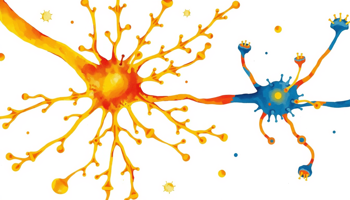
The answer lies at the synapse, the tiny gap between neurons where signals pass from one cell to the next. John Morrison and Mark Baxter at Mount Sinai published an influential review in Nature Reviews Neuroscience in 2012 that shifted the field's focus [1]. They showed that aging is fundamentally a synaptic phenomenon. Dendritic spines, the small protrusions on neurons where most excitatory synapses form, are selectively lost with age. In the prefrontal cortex of aged monkeys, thin spines, the most plastic and learning-related type, are reduced by nearly fifty percent [11]. The thicker, more stable mushroom spines are relatively preserved.
This matters because thin spines are the ones most involved in forming new associations. Lose them, and the brain keeps its old knowledge but struggles to encode new information. Sound familiar? That is exactly the pattern seen in normal aging: old memories intact, new learning harder.
The loss of dendritic spines also weakens long-term potentiation, or LTP, the cellular mechanism that strengthens connections between neurons during learning. Eric Kandel shared the Nobel Prize in 2000 for his work on the molecular basis of LTP. In aged brains, LTP is harder to induce and decays faster [12]. The machinery of learning is not broken. It is wearing down.
The Dopamine Decline
Beyond structural changes, the chemical environment of the brain shifts with age. The neurotransmitter most consistently linked to cognitive aging is dopamine.
Lars Bäckman, Lars Nyberg, and their colleagues at the Karolinska Institute proposed what they called the "correlative triad" in 2006: aging causes dopamine receptor loss, which causes cognitive decline [13]. A meta-analysis found that D1-like dopamine receptor availability correlates negatively with age at roughly r = -0.77, one of the strongest brain-behavior associations in all of cognitive neuroscience [14]. D2-like receptors decline at about five to ten percent per decade.
In 2024, Karalija and colleagues published the first major longitudinal confirmation of this triad. They followed older adults over five years, measuring striatal D2 receptor availability alongside working memory performance. The finding: individuals with the steepest dopamine receptor decline showed significant working-memory deterioration. Those whose dopamine levels remained more stable kept their cognitive edge [14].
What does this mean in practical terms? Dopamine is not just the "reward chemical" of popular science articles. It is the signal that tells the brain: this is worth paying attention to. This is worth remembering. When dopamine declines, the filter weakens. Irrelevant information gets in. Relevant information gets less emphasis. The brain becomes noisier.
The cholinergic system, centered on acetylcholine, also declines. Acetylcholine is critical for focused attention and memory encoding. Its reduction helps explain why older adults often say they need a quiet room to concentrate, while younger adults can study at a noisy coffee shop.

When Does Decline Actually Begin?
This question generates more scientific disagreement than almost any other in cognitive aging research.
Timothy Salthouse at the University of Virginia published a provocative paper in Neurobiology of Aging in 2009. Using large cross-sectional samples, he argued that fluid cognitive abilities, including some aspects of memory, begin declining as early as the late twenties [7]. Processing speed drops first. Reasoning follows. Memory is close behind.
K. Warner Schaie, whose Seattle Longitudinal Study tracked the same individuals over decades, fired back. He called Salthouse's findings a "cross-sectional fallacy." When you compare twenty-year-olds and sixty-year-olds at one point in time, you are not measuring aging. You are measuring generational differences in education, nutrition, health care, and technology. Schaie's longitudinal data showed meaningful decline starting closer to age sixty [15].
Who is right? Probably both, in different ways. Salthouse's own later work, using both cross-sectional and longitudinal designs with nearly 5,000 participants, confirmed that the two methods paint genuinely different pictures of the same phenomenon [16]. Cross-sectional data captures a real signal: processing speed does slow early. But longitudinal data captures a different truth: the functional impact of that slowing stays invisible for decades, masked by experience, strategy, and accumulated knowledge.
A reasonable synthesis: cognitive systems peak in the mid-twenties. Subtle changes in processing speed and associative memory may begin in the thirties and forties. Noticeable episodic memory changes emerge in the fifties and sixties. And acceleration happens after seventy, when hippocampal atrophy and the brain-memory coupling reach their strongest levels.

The SuperAgers Who Break Every Rule
Not everyone's brain follows the expected script. At Northwestern University, a research program now in its 25th year has been studying individuals over eighty whose memory performance matches that of adults twenty to thirty years younger. They call them SuperAgers [17].
The definition is precise. A SuperAger must be over eighty and must score at or above normal levels for fifty-to-sixty-year-olds on episodic memory tests. On the Rey Auditory Verbal Learning Test, that means recalling at least nine of fifteen words after a delay. Typical eighty-year-olds recall about five.
Sandra Weintraub, Marsel Mesulam, and their colleagues reported their 25-year findings in Alzheimer's and Dementia in 2025 [18]. Only about ten percent of their older participants qualified. What set SuperAgers apart? Their cortex was significantly thicker than that of same-age peers. Their anterior cingulate cortex, a region involved in attention and cognitive control, was actually thicker than that of younger adults. They lost brain volume at roughly half the rate of typical aging: about 1.06 percent per year versus 2.24 percent. They carried the APOE ε4 allele less often. And postmortem analysis showed roughly three times fewer tau tangles in their hippocampus.
SuperAgers are not simply people who got lucky with their genes. Many share lifestyle patterns: sustained social engagement, physical activity, and a willingness to challenge themselves mentally. But the biological basis is real. Their brains age more slowly at a measurable, structural level.
The Confusion Zone: Normal Aging, MCI, and Disease
One of the most important skills in medicine is distinguishing normal memory changes from early disease. The boundary is not always clear, and recent research has made the picture both more precise and more complex.
Normal age-related memory impairment means occasionally misplacing keys, blanking on a name, or forgetting why you walked into a room. These lapses do not interfere with daily life. Cues help: someone reminds you of the name and it comes back immediately.
Mild cognitive impairment, or MCI, is a middle ground. People with MCI show measurable cognitive decline beyond what is expected for their age, but they can still live independently. A 2022 meta-analysis of 66 studies involving 242,804 participants found MCI prevalence at about 15.6 percent among community-dwelling adults over fifty, rising steeply with age [19]. MCI is not dementia. But it increases the risk. Roughly eight to fifteen percent of MCI cases convert to dementia each year. However, a substantial fraction, up to 38 percent in the Mayo Clinic Study of Aging, revert to normal [20].
In 2024, Mayo Clinic researchers defined a new clinical syndrome: Limbic-predominant Amnestic Neurodegenerative Syndrome, or LANS. This condition looks like Alzheimer's at first glance: memory loss, hippocampal shrinkage, confusion. But it is driven by a different protein, TDP-43, not amyloid. LANS progresses more slowly and has a better prognosis than Alzheimer's disease. The clinical criteria, published in Brain Communications, now allow clinicians to distinguish LANS from Alzheimer's during a patient's lifetime, preventing inappropriate treatment [21].
Blood biomarkers are transforming early detection. Plasma phospho-tau 217, or p-tau217, can now detect Alzheimer's pathology with areas under the curve of 0.93 to 0.96, meaning it correctly classifies patients over ninety percent of the time [22]. The first Alzheimer's blood test received FDA clearance in 2025. This means the era of "we cannot tell you whether your memory problems are normal or pathological until it is too late" is ending.

What Can Be Changed
Perhaps the most powerful finding of the past decade is this: nearly half of all dementia cases may be preventable.
The 2024 Lancet Commission on Dementia Prevention, the most authoritative review in the field, identified fourteen modifiable risk factors that together account for about 45 percent of dementia cases worldwide [23]. The two largest single contributors: untreated hearing loss and high LDL cholesterol, each accounting for seven percent of cases. Less education contributes five percent. Social isolation, five percent. Depression, traumatic brain injury, and air pollution each contribute three percent.
Exercise is one of the best-studied protective factors. In 2011, Kirk Erickson and colleagues at the University of Pittsburgh published a landmark randomized controlled trial. Older adults who walked briskly three times per week for twelve months increased their anterior hippocampal volume by about two percent, effectively reversing one to two years of age-related shrinkage. Their spatial memory improved. Their blood levels of BDNF, brain-derived neurotrophic factor, a molecule that supports neuron growth and synaptic plasticity, increased [24].
Sleep matters too, and for reasons that go beyond rest. The glymphatic system, discovered by Maiken Nedergaard's lab in 2012, clears metabolic waste from the brain, including amyloid-beta and tau, the proteins that accumulate in Alzheimer's disease [25]. This clearance system is most active during slow-wave deep sleep, when interstitial space in the brain expands by up to sixty percent. Slow-wave sleep declines markedly with age [26]. The implication: as deep sleep diminishes, the brain's ability to wash away toxic proteins weakens. Poor sleep may not just be a consequence of brain aging. It may be a cause.
The MIND diet, developed by Martha Clare Morris at Rush University, combines elements of Mediterranean and DASH diets with emphasis on brain-protective foods: green leafy vegetables, berries, nuts, whole grains, fish, poultry, olive oil, and wine in moderation. In the Rush Memory and Aging Project, participants in the top third of MIND diet adherence showed cognitive decline equivalent to being 7.5 years younger than those in the bottom third [27]. A subsequent randomized trial published in the New England Journal of Medicine in 2023 found no significant difference over three years, though both groups improved, likely because the control group also upgraded its diet [28]. The observational evidence remains strong even if the trial was equivocal.
Hearing deserves special mention. The ACHIEVE trial, published in The Lancet in 2023, randomly assigned 977 adults aged 70 to 84 to hearing intervention or health education. In a prespecified subgroup of 238 higher-risk participants, hearing aids slowed cognitive decline by 48 percent over three years [29]. The mechanism likely involves keeping the brain socially and cognitively engaged. When hearing fails, conversations become exhausting, social participation drops, and the brain receives less stimulation.
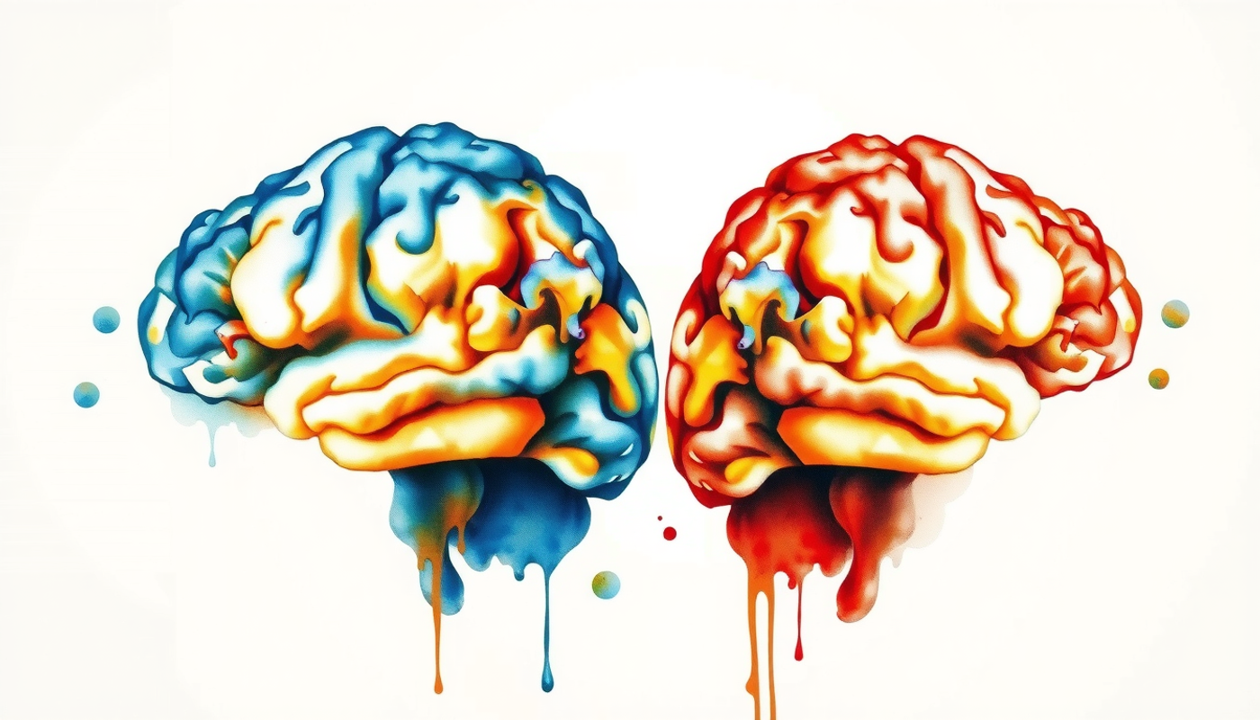
Sex, Hormones, and the Two-Thirds Problem
Almost two-thirds of Americans living with Alzheimer's disease are women. This disparity exceeds what greater female longevity alone can explain. Research from Lisa Mosconi at Weill Cornell Medicine has documented that the menopausal transition brings measurable brain changes: declines in estrogen-dependent memory, increased amyloid deposition, hippocampal volume loss, and reduced brain glucose metabolism [30].
Estrogen is neuroprotective. It supports hippocampal function, promotes synaptic plasticity, and modulates the cholinergic system. When estrogen levels plummet at menopause, the brain loses a protective shield. There is also a sex-gene interaction: a single copy of the APOE ε4 allele raises Alzheimer's risk by about 81 percent in women versus 27 percent in men [31].
Whether hormone replacement therapy can protect memory remains unresolved. The Women's Health Initiative Memory Study found unfavorable cognitive effects when hormone therapy was started after age 65 [32]. But the "critical window hypothesis" suggests that starting therapy near menopause, when receptors are still responsive, may be protective. Clinical trials are still testing this idea.
The practical implication for women: the standard risk factors (exercise, sleep, diet, hearing) apply equally, but women may benefit from additional attention to cardiovascular risk management during and after menopause.

The Frontiers: Senolytics, Glymphatics, and Blood Tests
The science of brain aging is moving fast. Three frontiers deserve attention.
Senolytic drugs aim to clear senescent cells, cells that have stopped dividing but refuse to die and instead secrete inflammatory molecules that damage their neighbors. In mouse models of tauopathy, intermittent treatment with dasatinib plus quercetin reduced tau accumulation, lowered neuroinflammation, preserved synaptic density, and restored cerebral blood flow [33]. The first Phase 1 human trials (SToMP-AD and STAMINA) have been completed, testing safety in older adults with early Alzheimer's markers. Results are preliminary but the concept is advancing.
The glymphatic system has moved from rodent discovery to human confirmation. Nedergaard's 2025 review in Science describes how neuromodulators oscillate in a coordinated rhythm during sleep, driving vasomotion that pumps cerebrospinal fluid through the brain's waste-clearance pathways [34]. Heart-rate variability during sleep may serve as a biomarker for glymphatic function, potentially giving clinicians a way to assess brain waste clearance with a simple wearable device.
Blood-based biomarkers are perhaps the nearest-term transformation. Plasma p-tau217 is already FDA-cleared. An analysis presented at AAIC 2024 showed it could predict amyloid positivity in cognitively unimpaired individuals with 79 to 86 percent accuracy [35]. This means routine screening for Alzheimer's pathology could become as simple as a blood draw, years before symptoms appear.
The Honest Counterarguments
Science moves forward by questioning its own findings. Several caveats deserve attention.
The Lancet Commission's 45 percent figure is a theoretical maximum, not a guaranteed achievable reduction. It assumes elimination of risk factors and accounts for overlap between them. In practice, no society has come close to eliminating all fourteen factors.
The exercise-hippocampus story is real but nuanced. In Erickson's trial, the stretching control group also improved on memory, suggesting that mechanisms beyond hippocampal volume growth contribute. Physical activity is unquestionably good for the brain. But the specific dose, type, and mechanism remain subjects of ongoing research.
The neurogenesis debate is genuinely unresolved. The studies by Boldrini and Sorrells used different tissue preparation methods and reached opposite conclusions about whether new neurons form in the adult hippocampus [6], [5]. This is not a settled question.
The cross-sectional versus longitudinal disagreement about when decline starts means that anyone who gives you a precise age ("decline begins at 27") is oversimplifying. The answer depends on which cognitive function, which measurement method, and which population you study.
And candidate-gene findings like BDNF Val66Met, KIBRA, and COMT have inconsistent replication records [36], [37]. The effects are real but small, and they vary across populations and study designs.

What This Means for Your Brain
The science of memory and aging, stripped to its essentials, says this: decline is real but not uniform, not inevitable, and not entirely out of your control.
In midlife, the highest-impact actions target cardiovascular health and sensory function. Treat hearing loss. Control blood pressure and cholesterol. Stay physically active. These are not vague wellness suggestions. They are interventions backed by large-scale epidemiological data showing that they collectively account for the largest modifiable fraction of dementia risk.
Across the lifespan, build cognitive reserve. Learning new skills, maintaining social connections, and engaging in cognitively demanding activities do not prevent neurons from dying (which is not what happens anyway). What they do is build redundant networks and alternative strategies that buffer against the structural losses that come with age.
Protect sleep. The glymphatic system's waste-clearance function depends on slow-wave sleep. Every night of poor sleep is a night of reduced brain cleaning. The compounding effect over decades may be substantial.
And pay attention to the signals. Normal forgetfulness, a missed name here or a misplaced object there, does not warrant panic. But progressive changes that interfere with daily life, that do not respond to cues, and that worry the people around you deserve professional evaluation. With p-tau217 blood tests now available, the distinction between normal aging and early pathology can be made earlier and more accurately than ever before.
The aging brain is not a broken brain. It is a brain that has traded speed for depth, quantity for quality, rapid encoding for accumulated wisdom. The challenge is protecting the systems that weaken while relying on the ones that do not.

Frequently Asked Questions
What is the difference between normal aging memory loss and dementia?
Normal age-related memory loss involves occasional forgetfulness that does not disrupt daily life. Forgetting where you left your keys but finding them later is typical. Dementia involves progressive decline that interferes with independence, such as getting lost in familiar places, repeating questions, or being unable to manage finances. A blood test for p-tau217 can now help distinguish the two.
At what age does memory start to decline?
This depends on the type of memory. Processing speed begins slowing in the late twenties. Episodic memory, the ability to recall specific events, typically shows noticeable changes in the fifties and sixties. Semantic memory, knowledge and vocabulary, often continues improving into the seventies. There is no single age at which all memory declines.
Can exercise really prevent memory loss?
A 2011 randomized controlled trial found that twelve months of aerobic walking increased hippocampal volume by about two percent in older adults. The Lancet Commission identified physical inactivity as one of fourteen modifiable risk factors for dementia. Exercise cannot guarantee prevention, but the evidence strongly supports it as one of the most effective brain-protective behaviors.
Why do women get Alzheimer's disease more often than men?
About 61 percent of Alzheimer's patients are women. Longevity plays a role, but estrogen loss at menopause also matters. Estrogen supports hippocampal function and synaptic plasticity. When it drops, the brain loses a protective factor. Women also show a stronger risk increase from carrying the APOE epsilon-4 gene variant compared to men.
What is a SuperAger?
SuperAgers are individuals over 80 whose episodic memory performance matches that of adults 20 to 30 years younger. Research at Northwestern University found they have thicker cortex, slower brain volume loss, fewer tau tangles, and lower rates of the APOE epsilon-4 allele. About 10 percent of adults over 80 meet SuperAger criteria.


