Introduction
Look up from this screen. Whatever your eyes land on, you already know what it is. There was no moment of effort, no sense of a decision being made, no felt gap between seeing and knowing. That absence of effort is the whole problem. It hides one of the most demanding computations your nervous system performs, and it has made visual pattern recognition unusually hard to measure. Photons hit the back of your eye. Roughly a tenth of a second later, cells deep in your temporal lobe are firing in a pattern that says dog or chair or the face of someone you have not seen in years. Nobody has fully explained the algorithm in between.
Search for how fast this happens and you will find five different answers. Thirteen milliseconds. One hundred. One hundred and twenty. One hundred and fifty. One hundred and ninety. Popular science articles quote whichever one they found first, usually without saying what it measures. The numbers look like a contradiction. They are not. They are five stopwatches clicked at five different moments in the same cascade, and once you know which moment each one marks, the disagreement dissolves and something much more interesting appears underneath: a precise, testable account of how a visual system turns light into meaning.
This article follows that cascade from the retina to the temporal lobe. It covers the classic experiment that gave us the 150 millisecond figure, the theories that tried to explain object recognition and mostly failed, the patients whose brain damage revealed the machinery by breaking it, the recent finding that a single forward sweep is not always enough, and the ongoing argument about whether the artificial networks that now predict brain activity are actually seeing anything at all.

The Experiment That Set the Clock
The number in this article's title comes from a single page and a half published in Nature in June 1996.
Simon Thorpe, Denis Fize and Catherine Marlot at the Centre de Recherche Cerveau et Cognition in Toulouse were trying to solve a measurement problem that had bothered vision science for decades. You can time how fast someone recognizes something by asking them to press a button. But a button press bundles two things together: the visual processing you care about, and the motor execution you do not. Reaction time is always an overestimate, and nobody knew by how much.
Their solution was to skip the muscles entirely and read the brain.
Subjects saw photographs they had never seen before, flashed for just 20 milliseconds. The task was a go/no-go categorization: press if the image contains an animal, withhold if it does not. Because the images were novel and natural, no low-level trick could solve them. A fox in undergrowth and a rock in undergrowth share almost every simple visual statistic. Meanwhile the researchers recorded event-related potentials from the scalp.
What they found was a frontal negativity specific to the no-go trials, developing roughly 150 milliseconds after the image appeared [1]. That signal is the fingerprint of a decision. The brain had already sorted animal from non-animal before any motor command was issued. Their conclusion was blunt: the visual processing required for this demanding task is finished in under 150 milliseconds.
Consider what has to fit inside that window. Light must be transduced by photoreceptors. The signal must cross the retina, travel the optic nerve, pass through a thalamic relay, enter primary visual cortex, and climb four or five further cortical stages, each adding complexity to the representation, until some population of neurons encodes an abstract category that generalizes across every fox, dog and beetle the subject has never seen before. In 150 milliseconds. With roughly ten milliseconds of processing available per cortical stage, there is barely time for each neuron in the chain to fire more than once.
Thorpe's group later showed the system is even more parallel than that. In a rapid animal categorization task, subjects responded as fast to two simultaneously presented natural images as to one [8]. High-level object representations were being accessed without sequential focal attention, in parallel, across the visual field.
Five Stopwatches, One Cascade
Here is the reconciliation. Each famous number marks a different event.
The 13 millisecond figure comes from Mary Potter's laboratory at MIT. Her team presented streams of six or twelve pictures with no gap between them, at rates from 13 to 80 milliseconds per picture, and asked subjects to detect a target named either just before or just after the sequence [2]. Detection stayed significantly above chance at every duration. This is not a claim that recognition finishes in 13 milliseconds. It is a claim about how briefly a picture can appear and still leave a usable trace, even when a new picture arrives immediately on top of it. The tradition goes back to Potter's own work in the 1970s on short-term conceptual memory for pictures [7].
The 100 and 120 millisecond figures come from the eyes rather than the hands. Holle Kirchner and Simon Thorpe flashed two scenes simultaneously, one in each visual hemifield, and asked subjects to look at the side containing an animal. Reliable saccades were launched in as little as 120 milliseconds, and low-level image differences could not explain them [5]. Sébastien Crouzet then ran the same design with faces and found the earliest reliable saccades at 100 to 110 milliseconds, with mean reaction times around 140 [4]. These very fast saccades were not entirely under instructional control. When faces were paired with vehicles, the eyes drifted toward faces even when subjects were told to target the vehicles.
That last detail deserves a moment. Something in the visual system is running ahead of your intentions.
The 150 millisecond figure is the neural decision signature described above. The 190 millisecond figure is the newest of the five. A 2024 EEG study from Yalda Mohsenzadeh's group at Western University used multivariate pattern analysis on naturalistic images in rapid sequences and separated two things that older work had blurred together. Gist perception, meaning partial conscious perception of what kind of thing is there, was decodable at roughly 120 milliseconds through feedforward mechanisms. Full object identification, meaning conscious perception of the specific image, resolved at roughly 190 milliseconds, with the processes for recognized and unrecognized targets diverging around 180 milliseconds in a way that implicates feedback [6].
So the five numbers line up like this: 13 is about exposure, 100 and 120 are about the eyes, 150 is about the decision, and 190 is about awareness of the specific thing.
The Asterisk on Thirteen Milliseconds
The 13 millisecond result is the one that travels furthest and gets quoted most loosely. It also has a serious challenge attached, and any honest treatment has to carry both.
In 2016, John Maguire and Piers Howe at the University of Melbourne pointed out a confound. In Potter's streams, each picture is supposed to mask the one before it. But natural photographs sometimes contain regions with no high-contrast edges. Where that happens, masking may be incomplete, and iconic memory of parts of the target picture could persist past the nominal presentation time, effectively giving the visual system longer than 13 milliseconds to work with.
They reran the study with four different mask types. With adequate masking, no evidence emerged that observers could detect a named target picture, even at 27 milliseconds per image [3]. Their conclusion was appropriately cautious: they could not rule out the possibility that feedback processing is necessary for individual pictures to be recognized.
This is not a debunking. It is a dispute about masking adequacy, and it remains unresolved. But it changes what the number means. Thirteen milliseconds is a headline that may be measuring the persistence of the visual buffer as much as the speed of recognition, which connects it directly to the brief store that George Sperling characterized in 1960 when he showed that a flashed array leaves behind far more information than subjects can report before it fades [17]. Anyone writing about the speed of vision should quote 13 milliseconds with the asterisk attached, or not quote it at all. If you want the fuller story of that fading buffer, it is covered separately in the quarter second most people never notice.

The Assembly Line
To understand why 150 milliseconds is remarkable rather than merely fast, you need to know how many stations the signal passes through.
The best single dataset comes from Matthew Schmolesky and colleagues, published in the Journal of Neurophysiology in 1998. They measured onset latencies of single-unit responses to flashed stimuli across the lateral geniculate nucleus and cortical areas V1, V2, V3, V4, MT, MST and the frontal eye field in individual anesthetized macaques, using identical procedures in each area, often in the same animal [9]. That methodological consistency is why the study is still cited nearly thirty years later. Most latency comparisons pool numbers from different labs using different anesthetics and different stimuli, which makes them close to meaningless.
Their headline findings: magnocellular geniculate layers led parvocellular layers by an average of 17 milliseconds. V1 responded before any other cortical area. A second wave arrived concurrently in V3, MT, MST and the frontal eye field. Latencies in V2 and V4 were progressively later and more broadly distributed.
Three warnings belong with this table, and competitors almost never give them.
First, these are macaque numbers, recorded under anesthesia. Human latencies run longer, on the order of 1.3 to 1.6 times, because human brains are bigger and the axons are longer. Anyone quoting 66 milliseconds for human V1 is quietly borrowing a monkey's brain.
Second, onset latency, peak latency and decoding latency are three different measurements and they give three different answers for the same area. Onset is when the first spikes appear above baseline. Peak is when firing is maximal. Decoding latency is when a classifier reading a population of neurons can tell one stimulus from another. They can differ by tens of milliseconds. A number without its metric is not a number.
Third, these are onset latencies for flashed stimuli in an anesthetized preparation, which is a deliberately simple case. Latencies shift with contrast, attention and stimulus complexity.
The V1 entry traces back to the single most durable result in this field. In 1962 David Hubel and Torsten Wiesel showed that cells in cat striate cortex respond to oriented edges, that simple and complex cells form a functional hierarchy, and that the cortex is organized into columns of shared orientation preference [10]. Everything built since, including the artificial networks discussed later, inherits that architecture.
At the top of the chain sits inferotemporal cortex. James DiCarlo, Davide Zoccolan and Nicole Rust summarized the consensus in 2012: core object recognition, the ability to recognize objects rapidly despite substantial variation in appearance, is solved by a cascade of reflexive, largely feedforward computations culminating in a powerful representation in IT [11]. The word doing the work there is largely. We will come back to it.
How good is that representation? Chou Hung, Gabriel Kreiman, Tomaso Poggio and DiCarlo answered in 2005. Reading out from roughly 100 randomly selected IT cells, over time windows as short as 12.5 milliseconds, they recovered accurate information about both object identity and category, generalizing across position and scale, even for novel objects [12]. A twelve millisecond glance at a hundred neurons is enough.
What the Human Brain Says
Macaque electrophysiology is precise but it is not us. Three human studies anchor the timeline.
Hesheng Liu, Yigal Agam, Joseph Madsen and Gabriel Kreiman recorded intracranial field potentials from 912 electrodes in 11 human patients undergoing epilepsy monitoring. They could decode object category from human visual cortex in single trials as early as 100 milliseconds after stimulus onset, and the decoding held up across depth rotation and scale changes [13]. Single trials, not averages. That matters, because averaging can manufacture apparent speed that no individual trial possesses.
Radoslaw Cichy, Dimitrios Pantazis and Aude Oliva combined magnetoencephalography and fMRI responses to 92 object images and used representational similarity analysis to link the two. Individual images were discriminated early by visual representations, while ordinate and superordinate category levels emerged relatively late. Early MEG responses corresponded to primary visual cortex and later MEG responses to inferotemporal cortex, giving a space-and-time-resolved picture of the first few hundred milliseconds of vision [14].
Leyla Isik, Ethan Meyers, Joel Leibo and Tomaso Poggio used MEG decoding to time the arrival of invariance itself. Object identity could be read out as early as 60 milliseconds. Size-invariant information appeared around 125 milliseconds and position-invariant information around 150 milliseconds, and both developed in stages, with tolerance to small transformations arriving before tolerance to large ones [15].
That staged arrival is the clearest evidence we have that invariance is built, not given. DiCarlo and David Cox framed the underlying problem memorably: every object produces a manifold of possible retinal images, and those manifolds are hopelessly tangled together in the retinal representation. The job of the ventral stream is to untangle them, one stage at a time, until a simple decision boundary can separate one object from another [16].

The Theories That Tried to Explain It
Long before anyone could time these events, psychologists proposed mechanisms. Most textbook and search-result treatments of visual pattern recognition still present these theories as a tidy progression that ends in a settled answer. The real picture is messier and more interesting.
Template matching was the simplest idea: store a copy of each known pattern, compare incoming input against the stored copies, pick the best match. It fails immediately. Rotate the object, change its size, move it, occlude it, and the match breaks. Storing a template for every possible appearance of every object requires impossible amounts of memory. Template matching survives today only as a baseline against which better ideas are measured.
Pandemonium, proposed by Oliver Selfridge in 1959, replaced whole-pattern matching with a hierarchy of feature detectors he called demons. Image demons pass raw input to feature demons, which shout in proportion to how strongly their feature is present. Cognitive demons listen for their own combinations of features and shout accordingly, and a decision demon picks the loudest. The specific implementation is long dead. The architecture is everywhere. Every hierarchical vision model since is a descendant.
Feature analysis got its physiological backing from Hubel and Wiesel. Real cells really do detect oriented edges, and they really are arranged hierarchically. This is the part of the classical picture that survived intact.
Prototype theory came from Eleanor Rosch in the mid-1970s. Categories are not defined by necessary and sufficient features but organized around central, typical examples, with membership graded by family resemblance [18], [19]. A robin is a better bird than a penguin, and reaction times reflect it. Prototype theory holds up well as an account of how categories are structured. It says less about the visual mechanism that gets you from photons to a category in the first place.
Recognition by components, Irving Biederman's 1987 theory, is the one that dominates search results. Objects are decomposed into a small alphabet of volumetric primitives called geons, roughly 36 of them, recovered from non-accidental properties of edges such as collinearity, curvature, symmetry, parallelism and cotermination. Because these properties survive changes in viewpoint, recognition should be viewpoint-invariant [20].
Marr and Nishihara's object-centered three-dimensional model representation, published in 1978, set the computational agenda for a generation [21]. Its influence on computer vision is hard to overstate even where its specific claims did not survive.
Geons Are Influential, Elegant, and Over-Taught
Biederman's theory deserves its place in the history of this field. It is also presented far more confidently than the evidence supports, and this gap is one of the clearest failures of existing content on visual pattern recognition.
The trouble arrived from two directions.
Michael Tarr and Heinrich Bülthoff tested whether human object recognition is better described by geon structural descriptions or by multiple stored views, and found consistent viewpoint-dependent costs: recognition slows as an object rotates away from a familiar view [22]. A strictly viewpoint-invariant theory does not predict that.
Then the neurons weighed in. Nikos Logothetis, Jonathan Pauls and Tomaso Poggio recorded from inferotemporal cortex in monkeys trained on novel objects and found cells tuned to particular views rather than to view-independent structural descriptions [23]. The brain appears to store families of views with learned tolerance between them, not abstract geometric skeletons.
Geons also struggle with the categories people care about most. Faces are not well described as arrangements of cylinders and wedges. Neither are trees, crumpled fabric, animals in motion, or most textured natural objects. The theory works best on exactly the class of rigid manufactured objects that the original demonstrations used.
The field moved toward hierarchical, learning-based, view-tolerant population coding. Maximilian Riesenhuber and Poggio's 1999 hierarchical model made that shift explicit, building invariance gradually through alternating selectivity and pooling operations rather than recovering a symbolic description [24]. Geons remain a landmark. They are not the current answer, and content that presents them as consensus is roughly forty years behind.
One Sweep, or Two?
Everything so far points one direction. The timing is too tight for anything but a single forward pass. Serre, Oliva and Poggio made the argument formally in 2007, showing that a feedforward architecture extending the Hubel and Wiesel simple-to-complex hierarchy predicted both the level and the pattern of human performance on a rapid masked animal versus non-animal categorization task [25].
But Victor Lamme and Pieter Roelfsema had already argued in 2000 that the feedforward sweep and recurrent processing offer genuinely distinct modes of vision. The sweep rapidly activates hardwired feature constellations across many areas. Recurrent processing, running through horizontal and feedback connections, handles perceptual grouping, figure-ground segregation and the binding that conscious perception seems to require [26].
For years this was a philosophical standoff. Then somebody found the images where it breaks.
Kohitij Kar, Jonas Kubilius, Kailyn Schmidt, Elias Issa and James DiCarlo reasoned that if recurrence matters, there should be images where primates beat feedforward-only networks. They used behavioral methods to hunt for hundreds of such challenge images. Then they recorded large-scale electrophysiology from IT. Behaviorally sufficient object identity solutions emerged roughly 30 milliseconds later for challenge images than for performance-matched control images, and those critical late-phase IT response patterns were poorly predicted by feedforward network activations [27].
Thirty milliseconds. That is the price of a hard image.
Kar and DiCarlo then asked where the extra signal comes from. Pharmacologically inactivating ventrolateral prefrontal cortex selectively degraded the late-solved images while leaving the early-solved ones intact, identifying prefrontal feedback as a critical recurrent node feeding back into IT [28].
Which images need the extra loop? The pattern is consistent across labs. Occlusion is the clearest trigger. Karim Rajaei and colleagues showed that recognizing partially occluded objects specifically recruits recurrent computation that feedforward models cannot account for [29]. Dean Wyatte, David Jilk and Randall O'Reilly found the same for objects degraded by noise and partial occlusion [30]. Clutter, unusual poses and low contrast push in the same direction.
This reframes the title of this article, and it should. One hundred and fifty milliseconds is the figure for an unoccluded object in a reasonably clean scene, viewed from a fairly ordinary angle. Put the same object behind a fence, or in a pile of other objects, or upside down, and the visual system takes longer because it has to run the loop. The headline number describes the easy case, which happens to be most cases, which is exactly why the system feels effortless.
When Recognition Breaks
The fastest route to understanding a machine is to find one that is broken in an informative way. Neuropsychology has provided several.
Visual agnosia is the loss of visual recognition with intact vision, intelligence and language. The classical division separates apperceptive agnosia, where the patient cannot build a coherent percept at all and cannot copy a drawing, from associative agnosia, where the percept forms and can be copied but carries no meaning.
Patient D.F. developed visual form agnosia after carbon monoxide poisoning. She could not report the orientation of a slot in front of her. She could not copy simple shapes. Yet when asked to post a card through that same slot, her hand rotated to the correct angle in flight, and when reaching for objects her grip aperture scaled correctly to their size. Perception was destroyed. Visually guided action was preserved. Melvyn Goodale and David Milner built the Two Visual Systems Hypothesis on this dissociation, proposing that the ventral stream constructs percepts while the dorsal stream converts vision into action [31]. A later review by Robert Whitwell, Milner and Goodale revisited D.F. with newer imaging and newer challenges, finding the core dissociation intact while the anatomical story turned out to be more complicated than originally described [32].
Patient H.J.A. showed a different fracture. Jane Riddoch and Glyn Humphreys documented a man with marked impairment in visual object recognition, good tactile object identification, and a preserved ability to copy drawings. His identification collapsed when figures overlapped, and depended heavily on how long he could look. His stored knowledge of objects was intact. The deficit was specifically in integrating form information into wholes [33]. They called it integrative agnosia.
Read those two cases together and you get an architecture. Vision for action separates from vision for recognition. Within recognition, feature extraction separates from integration, and integration separates from stored knowledge. Three dissociable stages, discovered by damage, matching the hierarchical cascade that electrophysiology independently described. Clinicians who train to read medical images are working the same machinery, which is why the way experts come to sort disease patterns into categories follows the same rules as ordinary object recognition.
The Face Area and the Argument That Never Ended
In 1997 Nancy Kanwisher, Josh McDermott and Marvin Chun reported an area in the fusiform gyrus that was significantly more active when subjects viewed faces than assorted common objects, in 12 of the 15 subjects tested. They ran further specificity tests within each individually defined region and named it the fusiform face area [34]. Kanwisher and Galit Yovel later reviewed the accumulated evidence for the region's specialization [35].
Then Isabel Gauthier proposed something uncomfortable. What if the FFA is not a face module but an expertise module that happens to be full of faces because faces are what everyone is expert at?
She trained people on artificial objects called greebles. Acquiring greeble expertise increased activation in right hemisphere face areas for upright greebles compared with inverted ones, and the same regions were more active in experts than novices during passive viewing [36]. She then extended the finding to real-world expertise, testing bird experts and car experts with fMRI. Homogeneous categories activated the FFA more than familiar objects, and right FFA and occipital face area showed significant expertise effects that tracked an independent behavioral measure of each participant's expertise [37].
The counterattack was immediate. Kanwisher argued for genuine domain specificity in face perception [38]. Elinor McKone, Kanwisher and Bradley Duchaine reviewed whether generic expertise could explain the special processing that faces receive, and concluded that the expertise effects were smaller and less consistent than the account required, while several signatures of face processing had no expertise analogue [39].
Where does it stand now? Softened, not settled. Almost everyone accepts that the FFA is strongly face-preferring. Almost everyone also accepts that expertise modulates it. What remains contested is whether the expertise modulation is the same phenomenon as face selectivity or a smaller effect riding on top of it. Anyone who tells you this was resolved in either direction is overstating the literature. The related question of why names detach so easily from faces we recognize instantly is a separate puzzle, explored in why faces stick when names do not.
Prevalence figures for developmental prosopagnosia carry a similar caution. The commonly cited number is 2 to 2.5 percent, traceable to Ingo Kennerknecht's German screening work [40]. In 2023, Joseph DeGutis and colleagues administered validated objective and subjective face recognition measures to an unselected web sample of 3,116 adults aged 18 to 55 and applied every diagnostic cutoff used in the previous 14 years. Estimated prevalence ranged from 0.64 to 5.42 percent depending on the cutoff, and the most commonly used researcher cutoffs produced a rate of 0.93 percent [41]. The familiar 2 percent is defensible as an upper-middle estimate. It is not a measured constant.
Machines That Predict the Ventral Stream
In 2014 something happened that reorganized this field.
Daniel Yamins, Ha Hong, Charles Cadieu, Ethan Solomon, Darren Seibert and DiCarlo searched a large space of biologically plausible hierarchical network models and discovered a strong correlation between a model's categorization performance and its ability to predict individual IT neural responses. They then took a high-performing network matching human performance on a range of recognition tasks and tested it against neural data it had never been fitted to. Its top layer predicted IT spiking responses to complex naturalistic images, and its penultimate layer predicted V4 [42].
Nobody told the network about brains. It learned to classify objects and ended up looking like a ventral stream.
Seyed-Mahdi Khaligh-Razavi and Nikolaus Kriegeskorte reached a convergent conclusion by a different route, showing that deep supervised models, but not unsupervised ones, came closest to explaining IT cortical representational geometry [43]. Martin Schrimpf and colleagues later formalized the comparison into Brain-Score, a public benchmark that scores any model against a battery of neural and behavioral datasets [44].
This is the strongest quantitative link between artificial and biological vision anyone has produced. It is also routinely oversold.
Where the Machines Stop Looking Like Us
Rajalingham and colleagues ran the most demanding version of the test. They collected over one million behavioral trials from 1,472 humans and five macaques, covering 2,400 images across 276 binary object discrimination tasks, then compared the behavioral signatures against feedforward convolutional networks. The networks accurately predicted primate patterns of object-level confusion, meaning which categories get mixed up with which. But at the level of individual images, the correspondence failed [45]. Humans and monkeys find the same specific images hard. The networks find different ones hard.
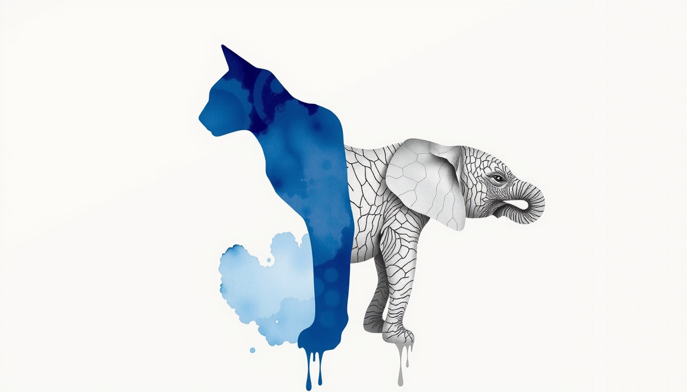
Robert Geirhos and colleagues then identified a systematic reason. ImageNet-trained networks are biased toward texture, while humans are biased toward shape. Show a cat silhouette filled with elephant skin, and the network says elephant while every human says cat [46]. Nicholas Baker, Hongjing Lu, Gennady Erlikhman and Philip Kellman found the same failure from another angle. Networks classified some silhouettes but showed no ability to classify glass figurines or outlines, and did not reliably distinguish an object's bounding contour from other edges [47]. Global shape, the cue human vision leans on hardest, is largely invisible to them.
Add adversarial examples, where imperceptible pixel changes flip a confident classification, and the picture gets uncomfortable. Jeffrey Bowers and thirteen coauthors made the case at length in Behavioral and Brain Sciences, arguing that benchmark prediction scores have been mistaken for mechanistic explanation, and that the field's evaluation methods are not designed to detect the ways these models diverge from human vision [48].
The fair summary is narrow and worth stating precisely. Deep networks are currently the best predictive models of ventral stream responses that exist. Predictive accuracy is not the same as mechanistic identity. And purely feedforward networks are missing the recurrent computations that Kar's challenge images showed the brain actually uses.
Half a Second Is Enough for a Radiologist
If the visual system builds category representations this fast, an obvious question follows. Does training change what it builds? Radiology gives the sharpest test available.
In 1975 Harold Kundel and Calvin Nodine showed ten radiologists a series of ten normal and ten abnormal chest films under two conditions: a 0.2 second flash, and unlimited viewing. With no time for visual search at all, overall accuracy reached 70 percent true positives. With free search it rose to 97 percent [49]. Seventy percent from a single glance, before the eyes could move even once.
Their interpretation shaped the field. Visual search begins with a global response that establishes content, detects gross deviations from normal, and then organizes the checking fixations that follow. Search is not a blind scan. It is a hypothesis being verified.
Karla Evans and colleagues rediscovered this with modern methods and pushed it further. Radiologists and cytologists rated briefly presented medical images for abnormality, then tried to localize the abnormality on a blank outline. Both groups performed above chance on detection [50]. Then came the result that makes the phenomenon undeniable. Evans, Tamara Miner Haygood, Julie Cooper, Anne-Marie Culpan and Jeremy Wolfe found that radiologists could discriminate normal from abnormal mammograms above chance after a half-second viewing, with a d-prime around 1, while remaining at chance in localizing the abnormality. The signal survived even when they viewed the mammogram of the opposite breast [51].
Detection without localization is the signature of a global gist signal rather than a found lesion. Trafton Drew, Evans, Melissa Võ, Francine Jacobson and Wolfe reviewed how this rapid global impression guides subsequent search through medical images [52]. The same principle underlies how students build durable visual memory for anatomical structures, where repeated structured exposure gradually turns effortful inspection into immediate recognition.
The Chessboard That Vanishes When You Shuffle It
Chess supplied the cleanest demonstration that perceptual expertise is real and, at the same time, the cleanest demonstration of its limits.
Adriaan de Groot found in the 1940s that after five seconds viewing a real game position, masters could reconstruct the board almost perfectly. William Chase and Herbert Simon formalized this in 1973 and gave it a mechanism: experts do not have larger memories, they have larger chunks. A master sees a castled king position as one unit where a novice sees six separate pieces [53].
Then they ran the control that made the finding matter. They shuffled the pieces into random configurations and repeated the test. The master's advantage nearly vanished.
That result is the honest heart of the expertise literature. What the master acquired was not memory capacity. It was a vocabulary of meaningful configurations, and outside the domain where those configurations occur, the advantage evaporates. Fernand Gobet and Simon extended chunking into template theory to explain how masters recall several boards at once, adding larger long-term retrieval structures while preserving the core finding that skill effects shrink dramatically for random positions [54].
Eyal Reingold, Neil Charness, Marc Pomplun and Dave Stampe then watched the eyes. Experts showed a substantially larger visual span for structured positions but not random ones, made fewer fixations, and often fixated on empty squares between related pieces rather than on the pieces themselves [55]. They were encoding relations in parallel, not pieces in sequence. This is the same fast, parallel, chunk-based encoding that the 150 millisecond literature describes, arriving through a completely different method. The broader pattern of how domain skill reshapes perception is covered in what separates experts from everyone else.
What Training Visual Pattern Recognition Can and Cannot Do
So perceptual expertise is trainable. The honest follow-up question is how far the gains travel, and here the literature is far less flattering than most popular accounts admit.
Philip Kellman and Patrick Garrigan developed perceptual learning modules, structured high-volume classification practice designed to accelerate the pattern-extraction process directly rather than teaching facts about it. Applied in medical image interpretation and mathematics, these produced substantial and durable improvements in accuracy and fluency [56]. Deliberate perceptual training works.
Now the limits.
Tomaso Poggio, Manfred Fahle and Shimon Edelman studied vernier hyperacuity, the ability to judge tiny spatial offsets. Learning was fast and required few examples. It was also stimulus-specific, and did not transfer between two only slightly different hyperacuity tasks [57]. Two tasks that look nearly identical to the person performing them are two separate skills to the visual system.
Fahle later reviewed the accumulated evidence on specificity versus generalization and described high specificity as the trademark of perceptual learning: gains are typically tied to the trained retinal location, orientation, spatial frequency and direction of motion, and frequently to the trained eye [58]. Train one eye and the other often learns nothing.
There is a genuine counterpoint. Lu-Qi Xiao, Jun-Yun Zhang, Rui Wang, Stanley Klein, Dennis Levi and Cong Yu developed a double-training paradigm, pairing conventional feature training at one location with irrelevant training at a second location, and achieved complete transfer of perceptual learning across retinal locations [59]. Specificity turns out to be partly a property of the training protocol rather than an absolute wall. But double training is a narrow, carefully engineered exception, not a general license.
The defensible conclusion is unglamorous and worth stating plainly. Training a radiologist on mammograms produces a radiologist who reads mammograms faster and more accurately. It does not produce a person with better vision in general, better attention in general, or better memory in general. The visual system optimizes locally. Claims of far transfer from perceptual training should be treated with the same skepticism as claims of far transfer from brain training games, and for the same reason: the mechanism that produces the gain is the same mechanism that confines it. This distinction between what feels like improvement and what has actually been learned shows up throughout the study literature, and it is the same trap described in recognition versus recall.

Conclusion
Start with the number in the title and hold it loosely.
One hundred and fifty milliseconds is a real measurement from a real experiment. Thorpe, Fize and Marlot flashed a photograph for 20 milliseconds and watched a frontal ERP separate animal from non-animal roughly 150 milliseconds later, before any muscle moved. That result has stood for three decades and the ventral stream latency data explain how it is possible: a chain of stations, each adding roughly 15 to 20 milliseconds and a little more abstraction, from oriented edges in V1 to whole objects in inferotemporal cortex.
But it is the figure for the easy case. Kar's challenge images need another 30 milliseconds and a loop through prefrontal cortex. Occluded objects need recurrence. Full conscious identification of a specific image resolves closer to 190 milliseconds. And the famous 13 millisecond result carries an unresolved masking objection that most articles quoting it never mention.
The theories that tried to explain this have fared unevenly. Feature detectors survived and became the foundation of both biological and artificial vision. Prototypes survived as an account of category structure. Templates died. Geons became a landmark that is still taught as though it were consensus, forty years after the field moved to view-based, learning-based population coding.
The patients taught us the most per case. D.F. separated seeing from acting. H.J.A. separated features from integration. Prosopagnosia separated faces from everything else, and the argument about whether that separation reflects a face module or an expertise module has been running for twenty-five years without resolution.
And the machines? They predict IT responses better than anything humans designed by hand, which is remarkable. They also classify by texture where we classify by shape, fail on the specific images we find easy, and lack the recurrent circuitry the brain demonstrably uses. Both facts are true at once.
The last thing worth keeping is the finding from chess. Shuffle the pieces at random and the grandmaster's advantage nearly disappears. Everything the expert gained was a vocabulary of structures that actually occur in the world. That is what visual pattern recognition is: not a general-purpose speed advantage, but a library of regularities built from experience, retrieved in a fraction of a second, and useless outside the world it was built from.
Which is exactly why it feels like nothing at all.
Frequently Asked Questions
How fast can the human brain recognize an object?
The neural signature of a category decision appears about 150 milliseconds after an image is shown, measured by Thorpe, Fize and Marlot in 1996 using event-related potentials. Full conscious identification of a specific image resolves closer to 190 milliseconds. Behavioral button presses take longer because they include motor execution.
Is it true that the brain processes an image in 13 milliseconds?
That figure comes from Potter and colleagues in 2014 and describes the shortest per-picture exposure in a rapid sequence that still allows above-chance detection. It is not the time recognition takes. Maguire and Howe reported in 2016 that with stronger masking the effect disappeared even at 27 milliseconds.
What is the ventral stream and why does it matter for recognition?
The ventral stream is the pathway running from primary visual cortex forward into the temporal lobe, sometimes called the what pathway. Signals climb through V1, V2 and V4 to inferotemporal cortex, gaining abstraction at each stage until neurons respond to whole objects regardless of position or size.
Are the classical theories of pattern recognition still accepted?
Partly. Hubel and Wiesel's feature detectors survived and underpin modern models. Rosch's prototype theory remains solid for category structure. Template matching is obsolete. Biederman's geon theory stays influential and widely taught, but viewpoint-dependence findings and view-tuned neurons moved the field toward view-based accounts.
Can visual pattern recognition be trained?
Yes, but the gains are narrow. Radiologists detect abnormality above chance from a half-second glimpse, and chess masters reconstruct real positions almost perfectly. Both advantages nearly vanish outside their domain. Perceptual learning research consistently finds specificity to the trained stimulus, location and often the trained eye.