

Introduction
A first-year medical student opens an anatomy atlas. Page after page of labeled structures. Brachial plexus. Celiac trunk. Circle of Willis. Three thousand names. Five thousand spatial relationships. And an exam in twelve weeks. The student reads. Highlights. Rereads. Then walks into the cadaver lab and freezes. Nothing looks like the textbook. The structures are not color-coded. They do not come with arrows. The kidney is buried behind fat. The nerve is tangled with vessels. The student who memorized every label cannot find a single structure in three dimensions.
This is not a failure of effort. It is a failure of method. Anatomy is a spatial discipline trapped in a two-dimensional curriculum. And the key to unlocking it has been hiding in plain sight inside the neuroscience of visual memory [1]
Visual memory in anatomy is not a study hack. It is the biological foundation of how spatial knowledge gets built, stored, and retrieved. The story stretches from George Sperling's 1960 flashcard experiments to Nobel Prize-winning discoveries about place cells in the hippocampus. From a 2023 study in Isfahan that measured the visual memory scores of 240 medical students, to body-painting experiments in Durham where students drew muscles on each other's skin and scored higher on exams. From Allan Paivio's dual-coding theory, which explains why pictures beat words in memory, to 2024 meta-analyses showing that virtual reality anatomy labs produce measurably better learning than textbooks [2].
This article tells that story. Not as a textbook chapter. As the narrative of a scientific question: why does the brain remember what it sees better than what it reads and what happens when medical education finally takes this seriously?
Ten Thousand Pictures and the Brain That Remembers Them All
How much can the visual brain actually hold?
More than anyone expected. In 1973, Lionel Standing at Bishop's University in Quebec ran what remains one of the most cited experiments in memory science. He showed participants up to 10,000 photographs, each for five seconds. Then he tested recognition. The result: participants correctly identified roughly 83% of the images they had seen even from a pool of thousands [3]. For smaller sets of a few hundred images, accuracy climbed above 90%.
The number seemed impossibly high. Skeptics wondered if participants were recognizing categories rather than specific images. In 2008, Timothy Brady, Talia Konkle, George Alvarez, and Aude Oliva at MIT settled the question [4]. They showed participants 2,500 object photographs over five and a half hours. Then they tested recognition with three increasingly difficult conditions. Could participants distinguish an object they had seen from an object of a completely different category? Accuracy: 92%. From a different object of the same category? 88%. From the same object in a different state or pose? 87%.
The takeaway was stunning. Visual long-term memory does not just store vague impressions. It stores detailed, specific representations of thousands of individual objects. The brain has a warehouse for images that makes any verbal memory system look tiny by comparison.
Now consider what this means for anatomy. A typical first-year course covers roughly 3,000 to 5,000 named structures [5]. If the visual memory system can store 10,000 detailed images, the raw capacity is there. The question is not whether the brain can hold enough anatomy. The question is whether students are encoding anatomy visually in the first place — or wasting their time with word lists that bypass the most powerful memory system they have.

Two Roads Through the Brain: The What and Where of Seeing
Visual information does not travel through the brain in a single stream. It splits.
In 1982, Leslie Ungerleider and Mortimer Mishkin at the National Institute of Mental Health published a landmark study based on lesion experiments in macaque monkeys [6]. They identified two distinct pathways leaving the primary visual cortex. One runs downward and forward through the temporal lobe. They called it the ventral stream. It answers the question "what am I looking at?" — recognizing objects, faces, colors, and identities. The other runs upward into the parietal lobe. They called it the dorsal stream. It answers "where is it?" — encoding spatial location, distance, and motion.
Melvyn Goodale and David Milner refined this in 1992, arguing that the dorsal stream is better described as a "how" pathway — specialized not just for location but for visually guided action: reaching, grasping, navigating [7].
For anatomy, this distinction matters enormously. Identifying that a structure is the brachial plexus engages the ventral pathway. Determining that its trunks pass posterior to the clavicle and superior to the first rib engages the dorsal pathway. A student who studies only from labeled diagrams trains the ventral stream. A student who manipulates a cadaver, rotates a 3D model, or sketches structures from memory trains both streams simultaneously. And both must integrate for the knowledge to become clinically useful.
The visual cortex itself — located in the occipital lobe at the back of the head — processes information through a hierarchy. Primary visual cortex (V1) detects edges and orientations. V2 handles contours and textures. V4 encodes color and simple shapes. And the inferior temporal cortex, at the bottom of the temporal lobe, handles whole-object recognition — which is why damage to this area can leave a person able to see but unable to recognize what they are seeing [8].
When a medical student looks at a cross-sectional CT image, the entire hierarchy activates. V1 processes the edges. V2 detects tissue boundaries. V4 distinguishes gray-white matter contrasts. Inferior temporal cortex recognizes the kidney. And the dorsal stream simultaneously locates it relative to the spine, the aorta, the psoas muscle. This is dual coding in action — not as abstract theory but as measurable neural computation.
The Hippocampus: Where Spatial Maps Become Anatomical Knowledge
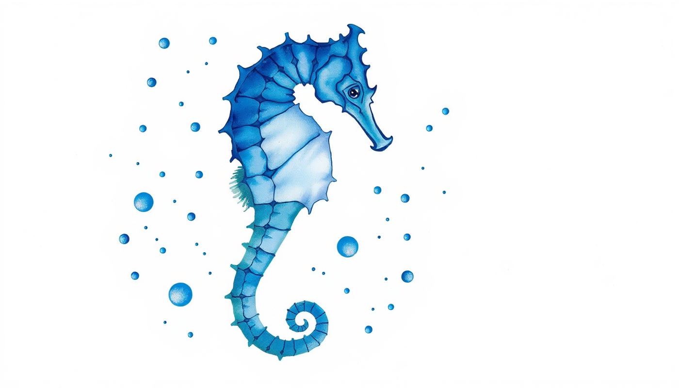
Deep inside each temporal lobe sits a curved structure shaped like a seahorse. This is the hippocampus — the brain's mapmaker and memory binder.
In 1971, John O'Keefe at University College London discovered something remarkable. He implanted tiny electrodes in the hippocampus of freely moving rats and found neurons that fired only when the animal occupied a specific location in space [9]. He called them place cells. Each place cell had its own preferred location. Together, thousands of place cells created an internal map of the environment.
Thirty-four years later, May-Britt and Edvard Moser at the Norwegian University of Science and Technology discovered grid cells in the medial entorhinal cortex — neurons whose firing locations formed perfect hexagonal grids, like invisible graph paper laid over the environment [10]. The 2014 Nobel Prize in Physiology or Medicine went to O'Keefe and the Mosers for discovering "cells that constitute a positioning system in the brain" [11].
What does this have to do with anatomy? More than most neuroscientists initially realized. In 2018, Jacob Bellmund and colleagues published a landmark paper in Science arguing that the hippocampal-entorhinal system does not just map physical space [12]. It maps conceptual spaces too. Grid-cell-like coding extends to abstract dimensions — categories, features, relationships. The brain uses the same spatial machinery to organize knowledge that it uses to navigate a room.
For anatomy, this means the hippocampus is doing double duty. When a student learns that the brachial plexus runs between the anterior and middle scalene muscles, the hippocampus is building a spatial map — encoding the plexus not just as a name but as a location relative to landmarks. When that student later encounters the same region in a cadaver or on a CT scan, the hippocampal map fires and recognition follows.
This is also why the method of loci — the ancient memory palace technique — works so well for anatomy. In 2003, Eleanor Maguire, Elizabeth Valentine, John Wilding, and Narinder Kapur at University College London scanned ten World Memory Championship competitors with fMRI [13]. The champions did not have unusually large brains or higher IQs. But during memorization, their brains showed significantly greater activation in the medial parietal cortex, retrosplenial cortex, and right posterior hippocampus — exactly the regions involved in spatial navigation. Nine of the ten champions confirmed using the method of loci.
For anatomy specifically, the body itself can serve as the palace. Students who trace the path of a nerve down their own arm while studying the brachial plexus are co-opting place cells and grid cells for anatomical content. The spatial memory system does not care whether the "space" is a building or a body. It maps both.
Why Pictures Beat Words: Dual Coding and the Picture Superiority Effect
Ask someone to remember a list of thirty words. Then ask a different person to remember thirty pictures. The pictures win. Every time.
This is the picture superiority effect, and it has been replicated so consistently since the 1960s that it qualifies as one of the most reliable phenomena in experimental psychology [14]. Roger Shepard showed it in 1967. Standing confirmed it at massive scale in 1973. Denis Nelson, Valerie Reed, and John Walling demonstrated it across multiple testing conditions in 1976.
The most influential explanation came from Allan Paivio at the University of Western Ontario. In 1971, Paivio proposed dual-coding theory: the mind processes information along two functionally independent but interconnected channels [15]. One channel handles verbal information — words, labels, linguistic descriptions. Paivio called these representations "logogens." The other handles nonverbal, imagistic information — shapes, spatial relationships, visual scenes. He called these "imagens."
The key insight: pictures automatically activate both channels. When you see a kidney, your visual system encodes the image (imagen) and your verbal system simultaneously generates the label "kidney" (logogen). But when you read the word "kidney," only the verbal system activates reliably. The visual system may or may not generate an image. Pictures get dual representation. Words get single representation. Dual representation means double the retrieval pathways, which means better recall.
James Clark and Paivio extended this to instructional design in a 1991 review in Educational Psychology Review [16]. Their conclusion: learning materials that combine text with relevant images consistently outperform text alone. Not because images are "prettier." Because they activate a second encoding system that text alone cannot reach.
What does this mean for anatomy? A student who reads "the brachial plexus passes between the anterior and middle scalene muscles" encodes a verbal trace. A student who simultaneously looks at a diagram or traces the path on their own body encodes a verbal trace and a visual-spatial trace. The second student has two pathways to recall. The first has one. Under exam pressure, two pathways are far more reliable than one.

The Isfahan Experiment: Measuring Visual Memory in Medical Students
Theory is one thing. Data is another.
In 2023, Amin Aspanani, Hosein Sadeqhi, and Athar Omid at Isfahan University of Medical Sciences published a study that put numbers on the relationship between visual memory and anatomy performance [1]. They recruited 240 medical and dental students — 148 medical, 85 dental — who were enrolled in anatomy courses. At the beginning of the semester, before any anatomy instruction, each student took two tests: the Jean-Louis Sellier visual memory test and ten items from the Gardner Spatial Intelligence Questionnaire.
Then they waited. At the end of the semester, they compared these baseline scores to the students' actual anatomy grades.
The results were clear. Medical students had significantly higher visual memory scores than dental students — mean 17.1 (±5.3) versus 14.3 (±4.6), with a p-value below 0.001. Pearson correlations showed a direct positive relationship between visual memory scores and anatomy course grades in both groups. Multiple linear regression confirmed it: visual memory predicted anatomy performance even when controlling for other variables.
The Isfahan study was not alone. In 2012, Lufler, Zumwalt, Romney, and Hoagland at Boston University administered the Vandenberg-Kuse Mental Rotations Test to 352 first-year medical students before their gross anatomy course [5]. Students in the top quartile of mental rotation ability were 2.1 to 2.2 times more likely to score above 90% on practical anatomy exams compared to the bottom quartile. Mental rotation scores also improved from pre- to post-course, suggesting that anatomy training itself sharpens spatial ability.
And in 2021, Roach and colleagues published a meta-analysis in Anatomical Sciences Education that pooled 15 studies [17]. Their finding: spatial ability consistently predicted anatomy performance with a small-to-moderate effect size. The strongest effects appeared on practical and drawing-based assessments — exactly the test formats that demand visual-spatial processing rather than verbal recognition.
The implication cuts both ways. Students with strong visual-spatial abilities have a measurable advantage in anatomy. But more importantly, spatial ability is trainable. Hoyek and colleagues showed that mental rotation training transferred to anatomy gains [18]. Vorstenbosch and colleagues at Maastricht University demonstrated that just one month of anatomy study improved mental rotation scores compared to controls [19]. Visual memory is not a fixed trait. It is a skill that anatomy education can — and should — deliberately build.
Drawing, Painting, and the Generation Effect
Reading about the brachial plexus is passive. Drawing it from memory is active. The difference is enormous.
In 2016, Jeffrey Wammes, Melissa Meade, and Myra Fernandes at the University of Waterloo published seven experiments comparing drawing to writing as a memory strategy [20]. Participants either wrote target words repeatedly or drew them for forty seconds. Drawn words were recalled at more than twice the rate of written words. The effect held even when controlling for elaborative encoding, visual imagery alone, and the picture superiority effect. Drawing was not just another way of seeing. It was a multimodal encoding act — combining visual, motor, semantic, and verbal codes simultaneously.
Fernandes and colleagues followed up in 2018 with a review in Current Directions in Psychological Science titled "The Surprisingly Powerful Influence of Drawing on Memory" [21]. They argued that drawing creates what they call "a seamless integration of semantic, visual, and motor codes" — the richest form of dual coding available without specialized equipment.
For anatomy specifically, the evidence is equally compelling. Body painting — where students use paint to draw anatomical structures directly on a peer's skin — has been tested repeatedly. Finn and McLachlan at Durham University analyzed 133 preclinical students across 24 focus groups and found that body painting produced stronger retention, color-linked memory, and higher engagement than traditional methods [22]. Jariyapong and colleagues confirmed improvements in surface anatomy knowledge for painted items [23]. And in 2025, Weeranantanapan and colleagues reported significant gains for muscular-system content with satisfaction ratings above 4.7 out of 5 [24].
Why does drawing work so well? The answer connects to another classic finding: the generation effect. Slamecka and Graf demonstrated in 1978 that self-generated information is remembered better than passively received information [25]. When a student sketches the branches of the celiac trunk from memory rather than tracing them from a diagram, the act of generation forces deeper processing. The student must retrieve spatial relationships, decide proportions, and correct errors — all of which strengthen the memory trace. Robert Bjork calls this a desirable difficulty — a condition that slows learning in the moment but dramatically improves long-term retention [26].
Spaced Retrieval: The Memory Schedule That Matches the Forgetting Curve
Visual encoding alone is not enough. Without scheduled review, even vivid memories decay.
In 1885, Hermann Ebbinghaus published the first quantitative measurement of forgetting [27]. His curve showed that roughly 70% of newly learned material disappears within 24 hours without review. Murre and Dros replicated and confirmed these numbers in 2015. The forgetting curve is not a metaphor. It is a measurable biological process.
Spaced repetition — reviewing material at gradually increasing intervals — directly attacks the shape of this curve. And when combined with visual learning, the effects multiply. B. Price Kerfoot at Harvard Medical School ran a series of randomized controlled trials testing spaced education in medical training. In 2010, he showed that web-based spaced quizzes improved long-term retention of histopathology knowledge among urology residents [28]. A follow-up study demonstrated that adaptive spacing — adjusting intervals based on individual performance — improved learning efficiency by 38% [29].
A 2024 systematic review and meta-analysis published in the Journal of Medical Internet Research pooled evidence from multiple trials of spaced digital education for health professionals [30]. The pooled effect on knowledge retention was significant (standardized mean difference = 0.38, 95% CI 0.10-0.65).
Image-occlusion flashcards represent the natural intersection of visual learning and spaced retrieval. In this format, anatomical diagrams are presented with labels hidden behind colored boxes. The student must actively recall what lies beneath — activating the testing effect that Roediger and Karpicke showed in 2006 to be one of the most potent learning strategies known [31]. Each successful retrieval strengthens the visual-spatial trace. Each failure signals the spacing algorithm to present the card sooner. The result is a self-correcting visual learning system that matches retrieval practice to individual forgetting rates.
Virtual Anatomy: What 24 Randomized Trials Actually Show
Three-dimensional anatomy tools have been around for over a decade. But do they actually work?
The best evidence comes from two independent meta-analyses published in 2024 in Anatomical Sciences Education. Salimi and colleagues analyzed 24 randomized controlled trials comparing VR-based anatomy instruction to traditional methods [2]. The pooled effect was moderate and statistically significant — standardized mean difference of 0.58 (95% CI 0.22 to 0.95, p < 0.01). But heterogeneity was high (I² = 87.4%), meaning the results varied widely across studies. Some trials found large benefits. Others found none.
García-Robles and colleagues conducted a parallel meta-analysis of immersive VR and augmented reality specifically [32]. They found significant pooled effects and consistently positive student perceptions, but again with substantial heterogeneity.
The most striking individual trial came from the TEACHANATOMY study published in Academic Medicine in 2025 [33]. Researchers randomized 48 students to learn cranial nerve anatomy either through a HoloLens 2 augmented reality module or through traditional textbook study. The AR group scored dramatically higher — median theoretical score 18.8 (IQR 16.6-20.0) versus 9.4 (IQR 7.7-11.3), p < 0.001. The effect was large. But the sample was small, and extrapolation should be cautious.
For 3D-printed anatomical models, Ye and colleagues meta-analyzed 13 RCTs in BMC Medical Education (2020) [34]. Standardized mean differences favoring 3D printing ranged from 0.37 (cardiac anatomy) to 2.01 (abdominal anatomy). Against cadavers, the SMD was 0.69. Against 2D images, it was 1.05.
The pattern across these studies is consistent but nuanced. Visual and immersive tools produce real learning gains. But the gains depend heavily on how the tools are used. Passive viewing of a 3D model produces minimal benefit. Active manipulation — rotating structures, stripping away layers, testing oneself — produces significant benefit. The technology is not the intervention. The pedagogy is. A 3D model used passively is just a prettier textbook.
When the Science Has Limits: What the Evidence Does Not Show
Intellectual honesty demands acknowledging gaps. And the evidence on visual memory in anatomy has several.
First, effect sizes are heterogeneous. Every meta-analysis in this field reports I² values above 80%, meaning the variation between studies is enormous. Sample sizes are often small — the TEACHANATOMY trial had just 48 participants. The Salimi VR meta-analysis pooled 24 trials but the individual studies ranged from 20 to 200 students, with different outcome measures, different anatomy topics, and different comparison conditions.
Second, the relationship between spatial ability and anatomy performance is real but modest. The Roach meta-analysis (2021) found small-to-moderate correlations, and the strongest effects appeared on practical exams. Multiple-choice exams — still the dominant assessment format in many medical schools — may underestimate visual memory's role because they rely heavily on verbal recognition rather than spatial recall [17].
Third, body-painting studies sometimes show non-significant overall differences despite significant gains on specific items. Jariyapong's 2016 study found improvements for painted surface-anatomy items but no statistically significant difference in overall MCQ scores [23]. Enthusiasm and engagement can confound objective outcomes.
Fourth, the neuroscience of place cells and grid cells comes primarily from rodent studies and human navigation tasks. The application to anatomy learning is theoretically well-motivated but lacks direct mechanistic experiments in medical education contexts. No one has implanted electrodes in a medical student's hippocampus during anatomy study. The inference that spatial memory circuits support anatomical learning is reasonable but indirect.
Fifth, cultural and curricular factors matter enormously. Cadaver availability, curriculum structure, hours of anatomy instruction, and assessment format all modulate every finding reported here. What works in a European gross anatomy lab may not transfer to a problem-based learning curriculum in Asia or a virtual-only program during a pandemic.
These caveats do not invalidate the evidence. They define its boundaries. The core finding — that visual-spatial encoding produces measurably better anatomy learning than verbal encoding alone — is supported by converging evidence from neuroscience, cognitive psychology, and medical education research. But the exact magnitude of the advantage, and the optimal way to achieve it, remain active areas of investigation.

From Lab to Clinic: How Visual Memory Transfers to Practice
Learning anatomy is not an end in itself. It is preparation for clinical medicine. And the visual skills built in anatomy transfer directly to radiology, surgery, and pathology.
Harold Kundel and Calvin Nodine demonstrated in the 1970s that experienced radiologists detect abnormalities on chest films within approximately 200 milliseconds — before their eyes have even completed a systematic scan [35]. This "holistic" or gist-based processing represents the expert endpoint of visual memory training that begins in anatomy.
Eye-tracking studies have mapped this expertise in detail. Van der Gijp and colleagues published a systematic review in Advances in Health Sciences Education (2017) describing the hallmark of expert radiological diagnosis: a global-focal search pattern [36]. Experts scan the entire image first, identify regions of interest, then focus. Novices jump immediately to salient features and miss surrounding pathology. The difference is not intelligence. It is trained visual pattern recognition — the downstream payoff of thousands of hours of visual anatomical learning.
In surgery, the connection is equally direct. Multiple studies have linked visuospatial ability and mental rotation to laparoscopic skill acquisition [37]. Kowalewski and colleagues (2021) showed that pre-existing visual-spatial ability predicts surgical performance during VR-simulator training, particularly on complex tasks. Surgeons navigate three-dimensional anatomy in real time under life-or-death conditions. The spatial maps they build during anatomy training are not academic exercises. They are clinical instruments.
Even in pathology — traditionally considered a microscopy discipline — visual pattern recognition drives diagnosis. Expert pathologists recognize tissue architecture at a glance. They distinguish normal from abnormal without counting cells or measuring structures. This expertise is visual, spatial, and built on a foundation of anatomical knowledge encoded through the visual memory system.

The Timeline: Key Discoveries in Visual Memory and Anatomy Learning
What This Means: Eight Evidence-Based Strategies
The science converges on a set of principles. Not tips. Not hacks. Principles grounded in how the brain actually encodes, stores, and retrieves spatial information.
First, convert text into images. Sketch pathways, vasculature, and cross-sections from memory. The drawing effect and the generation effect combine to produce encoding that is richer than any amount of rereading [21].
Second, use spatial mnemonics. The method of loci works because it co-opts the hippocampal spatial navigation system. For anatomy, the body itself is the palace. Trace the path of the brachial plexus down your own arm while studying it. Touch bony landmarks on your own body while learning muscle attachments.
Third, combine verbal and visual codes explicitly. Labeled diagrams with concise verbal summaries produce dual coding — the most robust form of memory encoding available [16].
Fourth, schedule retrieval rather than rereading. Image-occlusion flashcards with spaced repetition combine visual learning with the testing effect — two of the most potent learning strategies in cognitive psychology [31].
Fifth, interleave related topics. Mix gross anatomy with imaging anatomy with surface anatomy in a single session. Interleaving creates desirable difficulties that strengthen long-term retention.
Sixth, use 3D and immersive tools when available — but pair them with active retrieval. Passive viewing of a 3D model is little better than passive reading. Active manipulation followed by self-testing is transformative.
Seventh, train mental rotation explicitly. Visuospatial drills transfer to anatomy and surgical skill. The ability is not fixed — it improves with practice [18].
Eighth, use body painting and peer teaching when possible. Painting structures on a peer recruits motor, tactile, and visual codes simultaneously — the fullest embodiment of dual-coding theory.

Conclusion
Visual memory in anatomy is not a study preference. It is a biological reality. The human brain stores visual information with a capacity and detail that dwarfs verbal memory. It processes spatial relationships through dedicated neural circuits — the dorsal and ventral streams, the hippocampal place-and-grid-cell system, the posterior parietal cortex. And it encodes information most durably when multiple channels activate simultaneously — verbal plus visual, seeing plus doing, recognizing plus generating.
The evidence is consistent across levels of analysis. At the neural level, the visual cortex hierarchy, the hippocampal spatial mapping system, and the picture superiority effect all point in the same direction. At the behavioral level, studies from Isfahan to Boston to Waterloo show that visual-spatial ability predicts anatomy performance and that visual learning techniques produce measurable gains. At the clinical level, the visual expertise of radiologists, surgeons, and pathologists traces directly back to anatomical visual training.
And yet most anatomy curricula still rely heavily on lectures, labeled diagrams, and multiple-choice exams. The mismatch between how the brain learns spatial information and how medical schools teach it remains one of the most consequential gaps in medical education.
The science is not ambiguous. The brain learns anatomy best when it can see, touch, rotate, draw, and test itself in three dimensions. Every hour spent rereading a textbook page is an hour that could have been spent sketching from memory, manipulating a 3D model, or testing recall with image-occlusion cards. The visual memory system is the most powerful learning tool a medical student possesses. The question is whether education systems are ready to let students use it.
Frequently Asked Questions
How does visual memory differ from verbal memory in anatomy learning?
Visual memory stores spatial relationships, shapes, and three-dimensional structures through dedicated brain circuits including the ventral and dorsal visual streams. Verbal memory stores labels and sequences through language networks. Research shows anatomy students who engage both systems through dual coding recall significantly more than those relying on verbal study alone.
Can visual-spatial ability be trained or is it fixed?
Visual-spatial ability is trainable. Multiple studies show that mental rotation scores improve after anatomy coursework. Vorstenbosch and colleagues found that one month of anatomy study significantly improved spatial ability compared to controls. Hoyek and colleagues demonstrated that explicit mental rotation training transferred to anatomy performance gains.
Why does drawing from memory improve anatomy retention?
Drawing activates four encoding channels simultaneously: visual, motor, semantic, and verbal. Wammes and colleagues at the University of Waterloo found that drawing produced more than double the recall of writing. The act of generating a sketch from memory forces deeper processing and error correction, creating richer and more durable memory traces.
Does virtual reality actually improve anatomy learning outcomes?
A 2024 meta-analysis of 24 randomized controlled trials found a moderate, statistically significant effect of VR on anatomy knowledge compared to traditional methods. However, results varied widely across studies. Active manipulation of 3D models produces significant benefit, while passive viewing produces minimal gains. Pedagogy matters more than technology.
What is the picture superiority effect and why does it matter for medical students?
The picture superiority effect is the well-established finding that images are remembered better than words. Brady and colleagues showed humans recognize 92% of 2,500 previously seen objects. This matters for medical students because anatomy is inherently visual and spatial. Encoding structures as images rather than text labels exploits the brain's most powerful memory system.