

Introduction
A senior cardiologist glances at an ECG strip and says "anterior STEMI" before the intern has finished clipping it to the board. Two seconds. No checklist. No deliberation. Just a flicker of recognition and a diagnosis that will determine whether the patient lives or dies. How does the brain categorizes disease patterns this fast? And why does the same task take a third-year medical student several agonizing minutes of conscious reasoning?
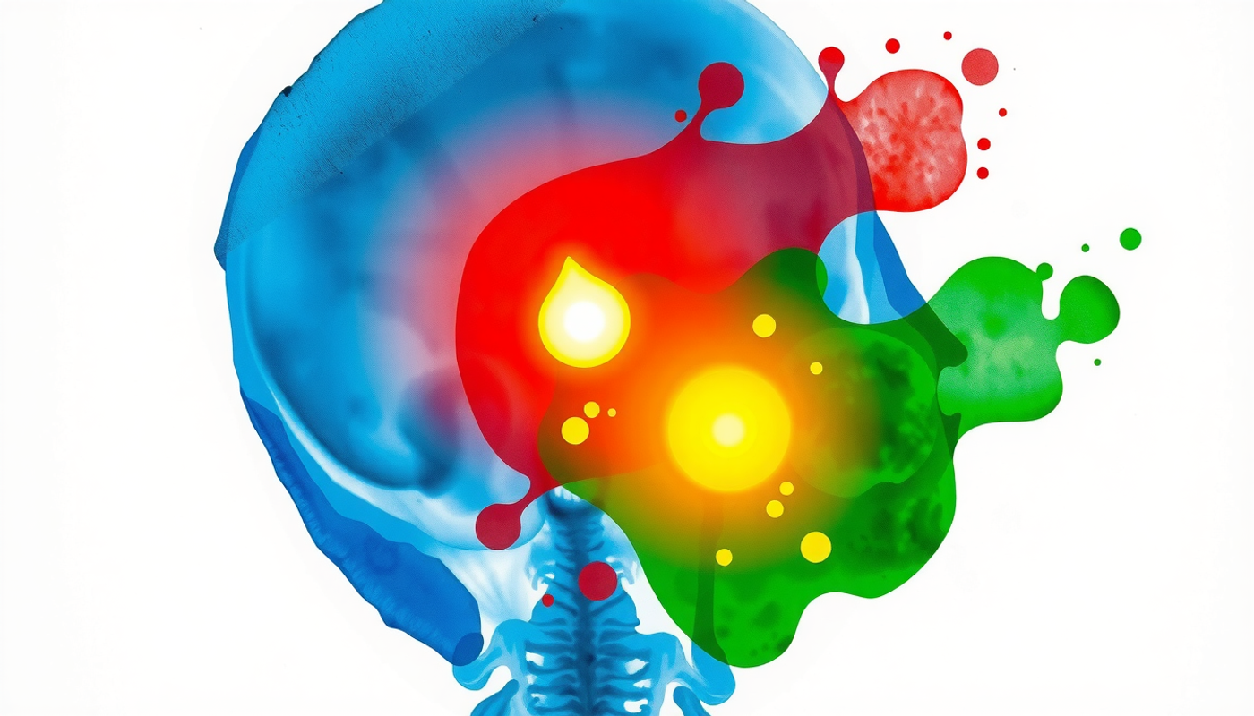
The answer is not simply "experience." It is a specific neural architecture that reorganizes itself over years of training — rewiring circuits in the basal ganglia, hippocampus, prefrontal cortex, and dopaminergic midbrain to transform slow, effortful analysis into rapid, automatic recognition [1]. This transformation follows the same computational rules the brain uses to categorize everything else — faces, birdsong, the smell of rain. But in medicine, the stakes are different. A misfire in pattern recognition does not mean mistaking a robin for a sparrow. It means missing a pulmonary embolism.
This is the story of how the brain learns to sort symptoms into diagnoses. It begins in a rat laboratory in the 1990s, passes through fMRI scanners in São Paulo and Rotterdam, and ends at the bedside of every physician who has ever felt the uncanny certainty of a diagnosis arriving before conscious thought catches up [2].
The Brain's Sorting Machine
Every diagnosis a physician makes is, at its core, an act of categorization. A bundle of noisy, incomplete signals — fever, cough, infiltrate on the chest X-ray — must be assigned to a stored category: "community-acquired pneumonia." This is the same computational problem the brain solves when it decides a four-legged animal is a dog rather than a cat. The mechanics are identical. The consequences are not.
The modern understanding of how the brain categorizes anything begins with Carol Seger and Earl Miller. Their 2010 review in the Annual Review of Neuroscience — titled simply "Category Learning in the Brain" — remains the field's reference text [1]. Seger and Miller argued that categorization is not a single process housed in a single brain region. It is distributed across multiple systems: the neocortex, the medial temporal lobe, the basal ganglia, and midbrain dopamine circuits. These systems interact through corticostriatal loops — anatomically distinct circuits that connect different cortical areas to different parts of the striatum — a structure buried deep inside the brain that acts as a switchboard for learned habits and decisions.
Three loops matter most for understanding medical expertise. The visual loop connects the visual cortex to the body of the caudate nucleus. It learns perceptual category boundaries — the kind a radiologist uses to spot a tumor on a chest CT. The executive loop connects the dorsolateral prefrontal cortex to the head of the caudate and the anterior cingulate. It supports rule-based categorization — the kind a medical student uses when she consciously runs through the diagnostic criteria for systemic lupus erythematosus. And the motor loop connects the motor cortex to the putamen. It supports reflexive, procedural categorization — the kind an experienced emergency physician uses when she recognizes a tension pneumothorax and reaches for the needle before finishing her sentence [1].
The critical insight from Seger and Miller is that these loops do not simply coexist. They compete. Early in learning, the executive loop dominates — everything is slow, deliberate, and conscious. With experience, control gradually shifts to the motor loop, and categorization becomes fast and automatic. This shift is not a metaphor. It is a measurable change in which brain regions activate during the same task.
Two Ways to Remember a Disease
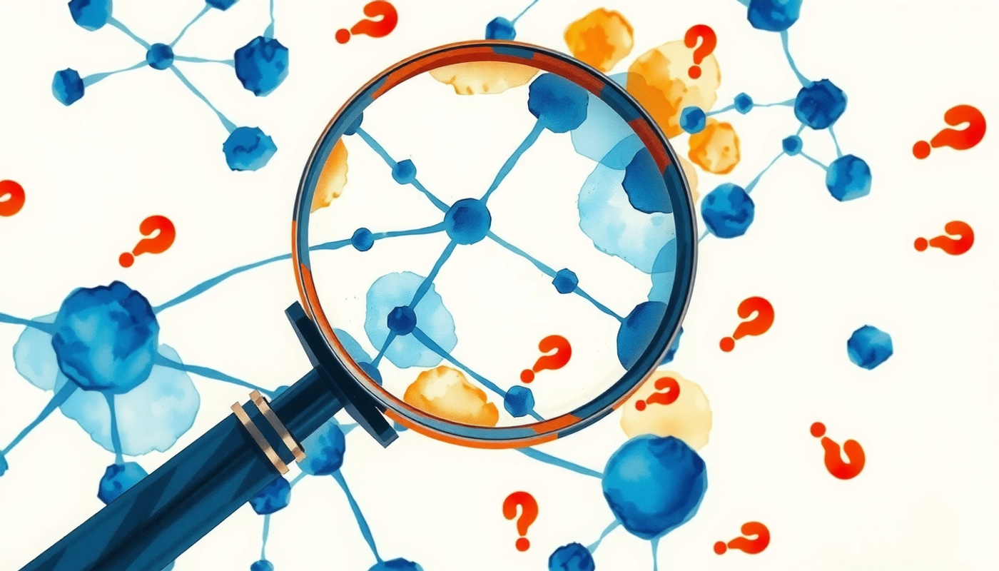
But where does the knowledge of diseases actually live in the brain? Cognitive psychology has debated this for decades, and the answer turns out to be: in two places at once.
One school says physicians store diseases as prototypes — averaged, idealized representations. "Cushing's syndrome" is a mental image of the textbook patient: moon face, buffalo hump, purple striae. When a new patient arrives, the brain compares her features to this prototype. The closer the match, the faster the diagnosis. The other school says physicians store diseases as exemplars — specific patients they have seen before. "Cushing's syndrome" is Mrs. Garcia from last Tuesday, or the memorable case from third-year clerkship. When a new patient arrives, the brain searches for the most similar past case.
In 2020, Caitlin Bowman, Yui Iwashita, and Dagmar Zeithamova at the University of Oregon published a study that settled the argument — or rather, showed that both sides were right [3]. Using fMRI, they tracked brain activity while participants learned new visual categories. The anterior hippocampus — the front part of the seahorse-shaped memory structure deep in the temporal lobe — and the ventromedial prefrontal cortex (vmPFC) — the patch of cortex just above the eye sockets — preferentially tracked prototype information. Meanwhile, the lateral occipital cortex, inferior frontal gyrus, and lateral parietal cortex tracked exemplar information.
What does this mean for medicine? When a physician thinks "this looks like a textbook case of Graves' disease," her vmPFC is doing the work. When she thinks "this reminds me of that patient from my intern year," her lateral cortex is retrieving an exemplar. Both systems operate simultaneously. Both contribute to the final diagnosis. And the balance between them shifts with expertise — experts rely more on prototypes for common diseases and more on exemplars for rare ones.
The vmPFC's involvement is particularly interesting. Bartra, McGuire, and Kable's coordinate-based meta-analysis of 206 fMRI experiments showed that this region encodes the subjective value of stimuli — essentially, how much something matters [4]. In clinical reasoning, this means the vmPFC is the region that says "this finding is diagnostically important" before conscious analysis has even begun.
Inside the Scanner: What Diagnosis Looks Like in the Brain
For decades, clinical reasoning was a black box. Researchers could ask physicians to think aloud, but they could not watch the process unfold inside the brain. That changed in 2012, when Steven Durning and his team at the Uniformed Services University of the Health Sciences began putting physicians inside fMRI scanners while they answered USMLE-style clinical vignettes [5].
The first studies established the basic anatomy. Diagnostic reasoning recruits a network that includes the dorsolateral prefrontal cortex — the brain's executive control center — the inferior parietal cortex, the anterior cingulate, and the caudate nucleus. Nothing surprising there. The surprise came when Durning compared board-certified internists to interns.
In a 2015 paper in Brain and Behavior, Durning's group showed that experts and novices shared the same core reasoning network [6]. Both groups activated the same regions. But experts showed something extra: activation in areas associated with automatized retrieval — the neural signature of knowledge so deeply encoded that it surfaces without effort. And in a follow-up by Hruska, Hecker, and colleagues, practicing gastroenterologists used fewer working-memory resources than novices for the same diagnostic tasks [7]. Same problem. Same answer. Less brain effort. This is neural efficiency — the hallmark of expertise.
But the most elegant demonstration came from São Paulo. In 2017, Melo, Scarpin, Amaro, and colleagues published a study in Scientific Reports that used time-resolved BOLD modeling — breaking the fMRI signal into 400-millisecond epochs — to watch diagnostic reasoning unfold in near-real-time [2]. Physicians read clinical vignettes word by word. When a highly diagnostic clue appeared — a symptom that strongly pointed to one diagnosis — something unexpected happened. Activity in the frontoparietal attention network decreased. The brain's monitoring system dialed down. The authors called this the neural signature of premature diagnostic closure — one of the most common causes of diagnostic error.
Think about what this means. The very mechanism that makes expert diagnosis fast and efficient — reducing monitoring when a pattern is recognized — is also the mechanism that makes it vulnerable to error. Speed and accuracy are not always friends.
Thinking Fast, Thinking Slow
In 2009, Pat Croskerry — an emergency physician and cognitive scientist at Dalhousie University in Halifax — published a paper that imported one of the most influential ideas from cognitive psychology into medicine [8]. The idea was dual-process theory, originally formulated by psychologists like Daniel Kahneman and Keith Stanovich: human cognition operates through two systems.
System 1 is fast. Intuitive. Automatic. It requires minimal cognitive effort. When an experienced dermatologist glances at a lesion and says "melanoma" in under a second, that is System 1. System 2 is slow. Deliberate. Analytical. It requires concentration and working memory. When a medical student works through the ABCDE criteria for melanoma — Asymmetry, Border, Color, Diameter, Evolution — that is System 2.
Croskerry argued that most of clinical practice runs on System 1. And most diagnostic errors originate there too. His 2013 paper in BMJ Quality & Safety, co-authored with Singhal and Mamede, catalogued more than 100 cognitive biases that affect clinical decisions [9]. Anchoring — locking onto an initial impression. Availability — overweighting diagnoses seen recently. Premature closure — accepting a diagnosis before it is verified. Each of these is a failure mode of System 1.
But here is where the story gets complicated. In 2020, van den Berg,";"; and colleagues at the University Medical Center Hamburg-Eppendorf published a study in Brain Communications that challenged the simple "System 1 bad, System 2 good" narrative [10]. They put sixteen experienced clinical neurologists — average twenty years of practice — inside an fMRI scanner and gave them two types of cases: straightforward ones and ambiguous ones that led to the same final diagnosis.
The straightforward cases activated the expected reasoning network. No surprise. But the ambiguous cases did not simply activate more of the same regions. Instead, they showed stronger connectivity between reasoning regions — the caudate, frontal cortex, and parietal cortex were talking to each other more intensely. Expert reasoning under uncertainty was not just "more System 2." It was a qualitatively different mode of processing — a tightly coupled network that could dynamically revise a provisional diagnosis as conflicting information arrived.
The lesson for medical students: expertise is not about choosing System 1 or System 2. It is about building a brain that can seamlessly switch between them and, when needed, run both at once.
The Scripts That Run Medicine
If System 1 is fast pattern recognition, what exactly is being recognized? The answer, according to forty years of research by Henk Schmidt, Remy Rikers, and their colleagues in Rotterdam, is illness scripts [11].
An illness script is a compact mental representation of a disease. It bundles three elements, originally described by Feltovich and Barrows in 1984 [12]: enabling conditions (who gets this disease — age, sex, risk factors, exposures), fault (what goes wrong pathophysiologically), and consequences (the resulting signs, symptoms, lab findings, and clinical course). An experienced physician's illness script for pulmonary embolism might look something like this: "postoperative patient or someone on a long flight (enabling condition) → clot forms in deep veins, embolizes to pulmonary arteries (fault) → sudden dyspnea, pleuritic chest pain, tachycardia, elevated D-dimer, wedge-shaped infiltrate on CT (consequences)."
The fascinating part is how these scripts develop. Schmidt and Rikers' encapsulation theory describes a four-stage process [11]. In the first stage, preclinical students accumulate detailed biomedical knowledge — the Krebs cycle, the coagulation cascade, the mechanisms of inflammation. This knowledge is stored as elaborate causal networks in semantic memory. In the second stage, during clinical clerkships, something remarkable happens. The biomedical details get "encapsulated" — compressed under higher-level clinical concepts. A student no longer thinks "factor V Leiden mutation causes resistance to activated protein C, which reduces the degradation of factor Va, leading to excessive thrombin generation." She thinks "hypercoagulable state." The mechanism is still accessible if needed, but it has been tucked away under a faster label.
In the third stage, these encapsulated concepts get integrated into illness scripts — rich, pre-compiled packages of knowledge organized around diseases rather than mechanisms. And in the fourth stage, with years of clinical experience, the scripts become populated with exemplars — specific patients whose faces and stories become part of the mental library.
This explains one of the most counterintuitive findings in medical education research: the "intermediate effect" [13]. When asked to recall clinical cases, senior medical students remember more biomedical details than expert physicians. Not because students know more, but because experts have encapsulated those details into higher-level categories. The details are still there — just not on the surface anymore.
The Dopamine Signal That Teaches Your Brain
Every diagnosis that turns out to be correct teaches the brain something. But how? The answer involves dopamine — the same neurotransmitter involved in motivation, pleasure, and addiction.
Wolfram Schultz's program of research, which earned him the 2017 Brain Prize, established that midbrain dopamine neurons signal reward prediction error [14]. When an outcome is better than expected, dopamine neurons fire. When an outcome matches expectations, they stay at baseline. When an outcome is worse than expected, they pause. This signal is not about pleasure. It is about learning. It tells the brain: "Update your model. Reality differed from prediction."
In category learning, this works like a graded teaching signal [15]. Every time a medical student makes a tentative diagnosis and gets feedback — from an attending, from a lab result, from the patient's clinical course — the basal ganglia receives a dopaminergic teaching signal. Correct diagnosis: positive prediction error, dopamine fires, the pattern gets strengthened. Incorrect diagnosis: negative prediction error, dopamine pauses, the pattern gets revised. Over thousands of cases, these signals sculpt the corticostriatal circuits that eventually enable automatic recognition.
The diagnostic "aha moment" — when a confusing case suddenly clicks into place — has the same neurochemical signature. Becker, Sommer, Kühn, and colleagues used ultra-high-field fMRI to image insight moments and found activation in the nucleus accumbens and ventral tegmental area — the core of the mesolimbic dopamine system [16]. The anterior superior temporal gyrus, first identified by Jung-Beeman and colleagues as the cortical locus of insight [17], also lit up. That satisfying "click" when a puzzling constellation of symptoms suddenly resolves into a diagnosis? That is dopamine reinforcing the pattern, making it more retrievable next time. This is why the difficulty of arriving at a diagnosis actuallystrengthens the memory effortful retrieval triggers stronger dopamine signaling and deeper encoding.
When the Brain Gets It Wrong
If pattern recognition is the engine of expert diagnosis, cognitive bias is the exhaust. And the most important study quantifying this problem was published in JAMA in 2010.
Sílvia Mamede's group at the Erasmus Medical Centre in Rotterdam recruited thirty-six internal medicine residents — eighteen first-year and eighteen second-year — and randomized them to a clever experimental design [18]. In the first phase, residents diagnosed a set of clinical cases. Some of these cases resembled — but were not identical to — cases they had seen before. In the second phase, they diagnosed a new set of "target" cases. The question: would seeing the similar-looking cases first bias their diagnoses of the target cases?
The answer was yes, but only for second-year residents. Their diagnostic accuracy dropped from 2.19 on a 0–4 scale (when they had not been primed) to 1.55 (when they had been primed with similar-looking cases). That is roughly a 29% relative decline. First-year residents showed a smaller, non-significant drop. This is the availability bias — a mental shortcut where the brain overweights diagnoses it has encountered recently.
But here is the critical finding. A second group of residents was asked to engage in "reflective reasoning" — explicitly considering alternative diagnoses and weighing evidence for and against their initial impression. Reflective reasoning recovered diagnostic accuracy to 2.03 for second-year residents — correcting roughly a third of the biased errors. The study demonstrated something actionable: structured reflection is a learnable cognitive skill that measurably reduces diagnostic error.
The emotional dimension matters too. Schmidt, Mamede, van den Berge, and colleagues showed that patients with disruptive behaviors — demanding, hostile, or non-compliant patients — reduced physicians' diagnostic accuracy by approximately 16% [19]. The mechanism was not that physicians disliked the patients (though they did). It was that managing the emotional reaction consumed cognitive resources — working memory that should have been processing clinical findings was instead processing frustration. Mamede's follow-up confirmed that the impairment was due to "doctors spending part of their mental resources on dealing with the difficult patients' behaviours, impeding adequate processing of clinical findings" [20].
Pat Croskerry's debiasing framework offers practical countermeasures [21]. His "cognitive forcing strategies" include: rule out the worst-case diagnosis first, consider the opposite of your initial impression, and force a "diagnostic time-out" before finalizing a diagnosis. These are not just good advice. They are structured interventions designed to engage System 2 when System 1 is likely to err.
The Eyes That See Disease
Some medical specialties depend almost entirely on visual pattern recognition. Radiology, dermatology, pathology, ECG interpretation — in these fields, the brain must categorize disease patterns from visual input alone, and the differences between novice and expert are dramatic.
In 2009, Harley, Pope, Villablanca, and Mumford compared first-year radiology residents, fourth-year residents, and practicing expert radiologists as they detected abnormalities in chest X-rays inside an fMRI scanner [22]. The result upended a long-standing assumption. The right fusiform face area (FFA) — a brain region famous for its role in recognizing faces — showed increased activity in expert radiologists compared to novices. The fusiform face area is not literally a "face" area. It is a region specialized for fine-grained within-category discrimination among visually similar objects. Gauthier and colleagues had already shown this in 2000: expertise with cars or birds recruits the same FFA region [23]. Harley's study showed that radiological expertise follows the same rule.
Even more striking: Bilalić and colleagues found that the FFA responded selectively to the fast, holistic mode of diagnostic processing — not to slow, deliberate search [24]. Expert radiologists can detect abnormalities in chest X-rays within two seconds, a process that resembles the holistic processing of faces.
How do expert eyes differ from novice eyes? Van der Gijp, Ravesloot, Jarodzka, and colleagues reviewed the eye-tracking literature across radiology and found a consistent pattern [25]. Expert search follows a "global-then-focal" model, originally described by Kundel and Nodine: experts first take in the whole image with a rapid global scan, form an initial impression, and then zoom in on suspicious regions. Novices skip the global scan and go straight to searching — a less efficient strategy that misses abnormalities visible only in context.
For ECG interpretation, Wood, Batt, Appelboam, and Wilson used eye-tracking on medical students and consultant emergency physicians [26]. Experts were roughly twice as fast to fixate the critical ECG leads. And a 2025 follow-up by Bortolotti and colleagues showed that expert readers interpreted ECGs in 108 seconds versus residents' 205 seconds — nearly double the speed — with significantly higher accuracy at every difficulty level [27].
From Student to Expert: How Years Reshape the Brain
The Dreyfus brothers — Hubert, a philosopher at UC Berkeley, and Stuart, an industrial engineer — proposed in 1980 that skill acquisition unfolds through five stages: novice, advanced beginner, competent, proficient, and expert [28]. Patricia Benner adapted this model for clinical nursing in 1984, and it has since become standard in medical education. At the novice stage, everything is rule-based and conscious. At the expert stage, performance is intuitive and situational — the expert "just knows" without being able to articulate the rules she is following.
But what happens to the brain during this transition? The fMRI evidence points to two changes. First, neural efficiency: experts activate fewer brain regions for the same task, as Hruska and colleagues demonstrated with gastroenterologists [7]. Second, network reorganization: experts do not just do less — they connect brain regions differently. The van den Berg 2020 study showed that expert neurologists showed stronger between-region connectivity, not just less local activation [10].
A resting-state fMRI study of radiology interns versus laypeople found that even baseline brain activity differs between people with and without visual diagnostic training [29]. Interns showed higher amplitude of low-frequency fluctuation (ALFF) in the right fusiform gyrus and left orbitofrontal cortex — changes correlated with their diagnostic performance. The brain had been reshaped not just during task performance, but at rest.
How long does this reshaping take? Anders Ericsson's influential 1993 paper in Psychological Review introduced the concept of "deliberate practice" — structured, feedback-rich training designed to target weaknesses [30]. Popular accounts simplified this to a "10,000-hour rule," but Ericsson himself rejected that number as misleading — it was a mean among elite violinists by age twenty, not a universal threshold. Macnamara and Maitra's 2019 replication found a smaller effect size for deliberate practice (η²=0.26 compared to Ericsson's original η²=0.70), suggesting that deliberate practice is necessary but not sufficient [31]. The defensible claim: thousands of hours of focused, feedback-rich clinical practice gradually reshape the corticostriatal circuits that underlie diagnostic expertise. But raw exposure alone is not enough.

The Science of Studying Smarter
The neuroscience of disease categorization is not just descriptive. It prescribes how to learn medicine more effectively.
Spaced repetition — reviewing material at increasing intervals rather than cramming — is the highest-evidence single intervention for medical education. Maye and colleagues' April 2026 meta-analysis in The Clinical Teacher analyzed fourteen studies covering 21,415 learners and found a standardized mean difference of 0.78 (95% CI 0.56–0.99) favoring spaced over massed study [32]. Mechanistically, spacing works because each retrieval event triggers hippocampal replay and cortical consolidation, gradually building the stable representations that will become illness scripts.
Interleaved practice — mixing different disease categories during study rather than studying one category at a time — is the second key intervention. Kornell and Bjork's 2008 study in Psychological Science demonstrated that interleaving improves long-term category discrimination even though it feels harder and slower [33]. The mechanism is discriminative contrast — by seeing pneumonia immediately followed by tuberculosis immediately followed by lung cancer, the brain is forced to identify the features that distinguish them. Blocking (studying all pneumonia cases, then all TB cases) never requires this discrimination, so it never builds it.
For radiology education specifically, a 2023 systematic review found that spaced learning, interleaving, and retrieval practice all improved diagnostic accuracy compared to traditional methods [34].
Retrieval practice itself is the third pillar. Roediger and Karpicke's 2006 study showed that the act of retrieving information is itself a memory-strengthening operation — more powerful than re-reading or even re-studying [35]. Larsen, Butler, and Roediger extended this to medical residents and found that repeated testing improved long-term retention of clinical knowledge compared to repeated study [36].
The practical prescription for medical students is clear. Study mixed-organ-system question banks (interleaving). Space study sessions across days and weeks rather than cramming (spacing). Test yourself actively rather than re-reading notes (retrieval practice). And after each practice case, ask: "What did I think first? Why? What disconfirming evidence did I underweight?" This last step — structured reflection — is Croskerry's debiasing routine, and it is the only intervention with replicated evidence for reducing cognitive bias in trainees [18].
What We Still Do Not Know
The picture painted so far is compelling but incomplete. Several caveats deserve honest acknowledgment.
First, fMRI samples in physician studies are small. Durning's expert sample was ten board-certified internists. Van den Berg's was sixteen neurologists. Wood's ECG study had ten students and ten consultants. These numbers are typical for expert-recruitment studies but produce wide confidence intervals. Replication is still incomplete.
Second, no fMRI study has directly imaged amygdala activation during physician diagnostic decisions. Claims linking the amygdala to affective diagnostic bias are theoretical extrapolations from Bechara and Damasio's somatic-marker work [37] and general affect-decision neuroscience, not direct evidence from clinical settings.
Third, dual-process theory is a useful descriptive framework, not a literal claim about two brain systems. Norman and Eva argued persuasively that the System 1/System 2 distinction is better understood as a continuum rather than a dichotomy [38]. And debiasing interventions have mixed empirical support — Norman and colleagues found limited evidence that teaching physicians about cognitive biases actually reduces diagnostic error [39].
Fourth, the prototype-versus-exemplar neural dissociation reported by Bowman and colleagues in 2020 is recent and not yet replicated at scale [3]. It is the cleanest current evidence for a dual-system account of categorization, but earlier work by Mack, Preston, and Love had argued for an exemplar-dominant brain representation [40]. The field remains active and the debate is not settled.
And fifth, the Mamede 2010 availability bias effect was statistically significant only for second-year residents, not first-years [18]. This suggests that bias susceptibility may actually increase during intermediate training before falling again with full expertise — what might be called the "experience-bias paradox." The most dangerous knowledge state may not be ignorance, but partial knowledge combined with premature confidence.
The Factory That Never Stops
The brain that categorizes disease patterns is not a filing cabinet. It is a factory — one that runs multiple production lines simultaneously, recalibrates its machinery with every case, and occasionally produces defective output that its own quality-control systems fail to catch.
The corticostriatal loops that Seger and Miller described [1] are the assembly lines. The illness scripts that Schmidt and Rikers mapped [11] are the products. The dopaminergic teaching signals that Schultz discovered [14] are the quality-control feedback loops. The dual-process architecture that Croskerry imported [8] is the decision about which production line to run. And the cognitive biases that Mamede quantified [18] are the defects that slip through.
What emerges from this research is a portrait of medical expertise that is both more impressive and more fragile than the traditional image of the wise physician. The expert brain is not simply a larger database. It is a fundamentally reorganized system — one where knowledge is compressed into scripts [12], perception is tuned by fusiform specialization [22], monitoring is calibrated by frontoparietal connectivity [10], and every correct diagnosis reinforces the circuit that produced it [14].
For the medical student reading this, the practical takeaway is concrete. The brain is already building your illness scripts — with every case you study, every question you answer, every diagnosis you get wrong and then correct. The neuroscience says you can accelerate this process: space your reviews, interleave your cases, test yourself actively, and reflect deliberately on your reasoning. Not because a textbook told you to. Because your corticostriatal loops, your hippocampus, and your dopamine neurons are listening — and they learn best when you make their job hard.

Frequently Asked Questions
What is clinical pattern recognition in medicine?
Clinical pattern recognition is the cognitive process by which physicians rapidly match a patient's symptoms, signs, and test results to stored mental representations of diseases. It relies on the brain's categorization systems in the basal ganglia, hippocampus, and prefrontal cortex and becomes faster and more automatic with clinical experience. Research shows that expert physicians can recognize common disease patterns in seconds.
How do illness scripts help doctors diagnose diseases?
Illness scripts are compact mental packages that bundle three elements of a disease: who gets it (enabling conditions), what goes wrong (pathophysiology), and what it looks like (signs and symptoms). Physicians build these scripts over years of training as detailed biomedical knowledge gets compressed into efficient clinical categories. They are the cognitive units that the brain retrieves during rapid pattern recognition.
What causes diagnostic errors in medicine?
Diagnostic errors most commonly result from cognitive biases in the fast, intuitive reasoning system. The most frequent contributors include premature diagnostic closure, availability bias from recently seen cases, and anchoring on an initial impression. Research by Mamede and colleagues showed that availability bias alone can reduce diagnostic accuracy by roughly 29 percent, though structured reflective reasoning can recover about one-third of those errors.
Can medical students improve their diagnostic pattern recognition?
Yes. Neuroscience research supports three evidence-based strategies for building stronger diagnostic pattern recognition: spaced repetition of clinical material, interleaved practice mixing different disease categories, and active retrieval testing rather than passive re-reading. A 2026 meta-analysis of over 21,000 learners found that spaced repetition produced a large effect size of 0.78 compared to massed study.
What brain regions are involved in medical diagnosis?
Medical diagnosis engages a distributed network including the dorsolateral prefrontal cortex for executive reasoning, the caudate nucleus in the basal ganglia for pattern matching, the hippocampus for memory retrieval, the ventromedial prefrontal cortex for evaluating diagnostic significance, and the frontoparietal attention network for monitoring information. Expert physicians show greater neural efficiency and stronger connectivity between these regions.


