Introduction
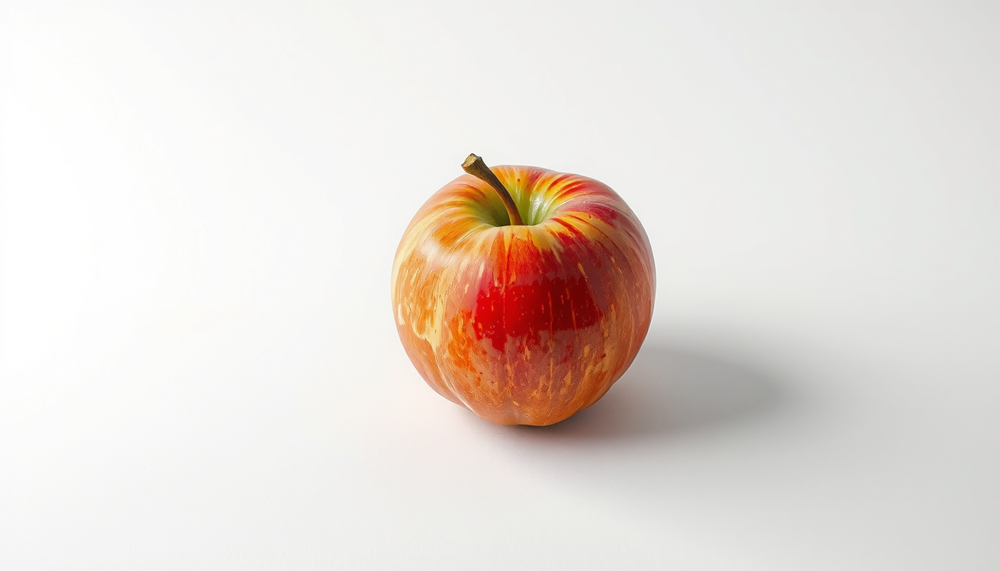
Close your eyes and think of an apple.
For most people something appears. A shape, a colour, maybe a shine on the skin. It might be faint or it might be sharp enough to count the freckles. Either way, something shows up.
For roughly one to four people in every hundred, nothing does. Not a dim outline. Not a shadow. Nothing at all. The knowledge is fully present. They can tell you an apple is round, that it is often red, that it has a stem and a core and a particular kind of crunch. They simply cannot see it. This is aphantasia, and the neurologist Adam Zeman gave it that name in 2015 after a decade of accidental discovery [1].
Here is the point that gets lost almost everywhere this subject is written about. Aphantasia does not mean bad memory. People with aphantasia recognise faces, hold down demanding careers, pass exams, learn languages, and recall their own lives. Some of them work at the very top of visual professions. What changes is not how much memory holds. What changes is what the act of remembering feels like, and which mental strategies do the work when the picture never arrives.
That distinction is where the science gets interesting, and where it gets messy. Over the last eight years, researchers have run drawing experiments, measured pupils, tracked sweat glands, and mapped brain lesions, all trying to answer a question that sounds simple and is not: if you take mental pictures out of a mind, what happens to memory? The answers so far do not agree with each other. Some studies find clear deficits. Others find almost nothing. Both sets of studies are well designed. Understanding why they disagree turns out to be more useful than picking a side.
This article follows that argument from a Victorian breakfast table to a 7-tesla scanner. It has real characters, unresolved fights, and a fair amount of evidence that overturns things everyone assumed were settled.
The Breakfast Table That Started Everything
In 1880 Francis Galton published a short paper in the journal Mind with the title Statistics of Mental Imagery [2]. He had sent a questionnaire to a hundred adult men, at least half of them prominent in science or other intellectual work, and to a group of schoolboys. The instruction was disarmingly domestic. Think of your breakfast table as you sat down to it this morning. Now rate the illumination of the image, its definition, and its colouring.
Galton expected variation. He did not expect the range he found.
Some respondents described mental scenes so detailed they could read the pattern on a plate. Others reported nothing whatsoever, and several were openly baffled that the question was being asked seriously. Galton noted that the people who most firmly denied having any imagery were often the ones who used visual language most fluently in conversation. They said things like "I picture it" while apparently picturing nothing.
He also drew a conclusion that stuck for over a century and turns out to be wrong. Galton suggested that scientists in particular were deficient in imagery, as though abstract thinking crowded out the pictures. In 2006 William Brewer and Marlene Schommer-Aikins went back to Galton's own published data, added a modern replication, and found no support for it [3]. Not a single scientist in the sample totally lacked imagery, and very few had genuinely feeble imagery. The founding claim of the field was a misreading of the founder's own numbers.
Galton's questionnaire also set a pattern that still causes trouble. He measured an invisible thing by asking people to describe it. Almost a century later, in 1973, David Marks formalised that approach into the Vividness of Visual Imagery Questionnaire [4]. Sixteen items, four scenarios, each rated from one to five, where five means no image at all and you merely know that you are thinking of the object. It became the standard instrument. It is still the standard instrument. And as we will see, it is also the single biggest reason researchers cannot agree on how common aphantasia is.
Then the topic went quiet for the better part of a hundred years.
The Man Who Lost His Mind's Eye
The modern story starts with a patient, not a theory.
In 2010 Zeman and colleagues published a case report in Neuropsychologia about a man they called MX [5]. He was in his sixties. A few days after a coronary angioplasty, his inner visual world switched off. His eyesight was unaffected. He could see the room, read a newspaper, recognise his family. But when he closed his eyes and tried to bring up a face or a place, there was nothing there.
The paper's title says what makes the case remarkable. Loss of imagery phenomenology with intact visuo-spatial task performance. MX scored close to the floor on imagery questionnaires. Then he sat down and performed normally on the standard laboratory tasks that psychologists had spent decades assuming required mental imagery. He could answer questions about the shapes of letters. He could do visual memory tests. He got the right answers without the pictures.
The brain scans made it stranger. When MX looked at photographs of famous faces, his fusiform activation looked like everyone else's. When he tried to imagine those same faces, the pattern changed. Control participants normally show posterior visual activity during imagery. In MX that posterior activity was reduced, while frontal activity rose. Something had changed in how his brain approached the task, not in whether it could do the task.
His dreams initially lost their visual content and then, over time, got it back.
MX was a single patient, which is worth saying plainly. But the case did something no theory could have. It made the phenomenon concrete and repeatable enough to write about, and it landed in front of the general public when the science writer Carl Zimmer covered it in Discover magazine. That is when Zeman's inbox started filling up.
Naming a Thing Changes Who Finds It
The people who wrote to Zeman after the Discover article had a different story from MX. They had not lost anything. They had never had it in the first place.
In 2015 Zeman, Michaela Dewar and Sergio Della Sala published a three-page paper in Cortex describing twenty-one of these correspondents [6]. They needed a word. A classicist friend suggested adapting Aristotle's term phantasia, the mind's eye, with the privative prefix. Congenital aphantasia.
The response was disproportionate to the paper. Three pages, and tens of thousands of people got in touch over the following years. Many described the same jolt of recognition: the discovery, often well into adulthood, that "picture it in your head" was not a figure of speech for everyone else. Zeman has since put the number of public contacts to his lab at around seventeen thousand [1].
That surge produced the largest early dataset. In 2020 Zeman and a large team published the Phantasia study in Cortex, drawing on questionnaire responses from roughly two thousand people with aphantasia and around two hundred with unusually vivid imagery, which they called hyperphantasia [7]. The scale was unprecedented. The recruitment method was also its main weakness, and the authors said so. Everyone in that sample had gone looking for the researchers after reading about the topic. That is not a random sample of the population, and any percentage drawn from it has to be treated accordingly.
Not everyone accepted the phenomenon at face value. In 2016 Stefania de Vito and Paolo Bartolomeo published a commentary in Cortex asking whether some cases might be psychogenic, a refusal or inability to engage with the task rather than an absence of the underlying capacity [8]. It was a fair challenge. Everything on the table at that point was self-report. If you want to establish that someone genuinely lacks an inner picture, asking them is not enough, because the thing you are asking about is only visible from the inside.
Answering that challenge took another six years and three very different experiments.
Each of those three experiments attacked the problem from a different angle. None of them asked anyone to describe their inner life.
Proving a Negative
The first came from Rebecca Keogh and Joel Pearson at the University of New South Wales, published in Cortex in 2018 under the title The blind mind [9].
The method exploits a quirk of vision called binocular rivalry. Show each eye a different image, say a red horizontal grating to one and a green vertical grating to the other, and the brain cannot fuse them. It flips between them. Which one you see first can be nudged by what you were imagining beforehand. In people with typical imagery, spending six seconds imagining the red horizontal pattern makes it more likely to win the rivalry contest. That priming effect is reliable and it does not depend on anyone reporting how vivid the image felt.
Keogh and Pearson ran fifteen people with aphantasia through the task and compared them against a pooled general-population sample of 209.
The general population primed strongly, well above the fifty percent chance level, with a t value of 10.96 and a p value below .001. The aphantasic group sat at chance. Their t value was 0.68 with a p of .51. A bootstrap analysis drawing a thousand random samples of fifteen from the general pool produced only one sample with a mean as low as the aphantasic group, giving a p of .001.
The obvious objection is that these participants were simply not trying. The study anticipated it. Mock rivalry trials, designed to catch response bias, showed no difference from chance for the aphantasic group either, which is what you would expect from people engaging honestly with a task that produced nothing to work with. A follow-up in 2024 extended the sample past fifty participants and found the same pattern [10].
One detail from that follow-up deserves more attention than it usually gets. Around twelve percent of aphantasic participants scored above sixty percent on the priming task. A minority appear to have something operating below the threshold of conscious experience. The group result is not the whole story.
The second experiment measured fear. In 2021 Marcus Wicken, Keogh and Pearson published in Proceedings of the Royal Society B a study on skin conductance, the same physiological arousal signal used in lie detection [11]. Participants read frightening first-person scenarios. Being chased. A plane losing altitude. In people with typical imagery, skin conductance climbed as the story unfolded. In twenty-two people with aphantasia, compared against twenty-four controls, it stayed flat.
Then came the control condition that makes the finding matter. A separate group, sixteen with aphantasia and fifteen without, viewed frightening photographs instead of reading text. Both groups responded normally. The physiological machinery for fear was intact. What was missing was the step where words are converted into an internal image vivid enough to frighten the body.
The third experiment measured pupils.
Your pupil constricts in bright light. It also constricts, slightly, when you merely imagine something bright. In 2022 Lachlan Kay, Keogh, Thomas Andrillon and Pearson published in eLife the study that closed the argument for most researchers [12]. Forty-two general-population participants and eighteen with aphantasia, the latter confirmed by a VVIQ score below 32 and binocular rivalry priming below sixty-five percent.
In the general population the imagery pupil response was overwhelming. An F value of 67.42 with a Bayes factor above ten to the power of ten. In the aphantasic group it was absent, F of 0.193 and a Bayes factor of 3.18 in favour of the null. The comparison between groups produced a Bayes factor above ten to the power of six.
And then the detail that settles the effort question for good. When aphantasic participants were asked to imagine four objects instead of one, their pupils dilated significantly with the increased cognitive load, F of 6.185 with a p of .02. They were working. The mental effort was measurable. The picture simply was not being produced.
Three independent laboratories, three unrelated physiological systems, one consistent result. The scepticism that de Vito and Bartolomeo raised in 2016 was reasonable at the time and has largely been answered. That does not mean the question of what aphantasia is has been answered, only the question of whether it is something.
Which brings us to memory, where the agreement stops.
The Drawing Experiment That Split the Field
Wilma Bainbridge runs a memory lab at the University of Chicago. Her speciality is a technique that sounds almost too simple to be rigorous: ask people to draw what they remember, then measure the drawings.
In 2021, working with Zoë Pounder, Alison Eardley and Chris Baker, she published in Cortex a study titled Quantifying aphantasia through drawing [13]. Sixty-one people with aphantasia and fifty-two with typical imagery studied photographs of three real-world rooms. Then they drew the rooms from memory. Later they copied the same photographs while looking at them, which controls for drawing ability. The drawings were scored by 2,795 online raters on two separate dimensions: how much object detail was present, and how spatially accurate the layout was.
The results split cleanly down the middle, and that split is the most important finding in this entire field.
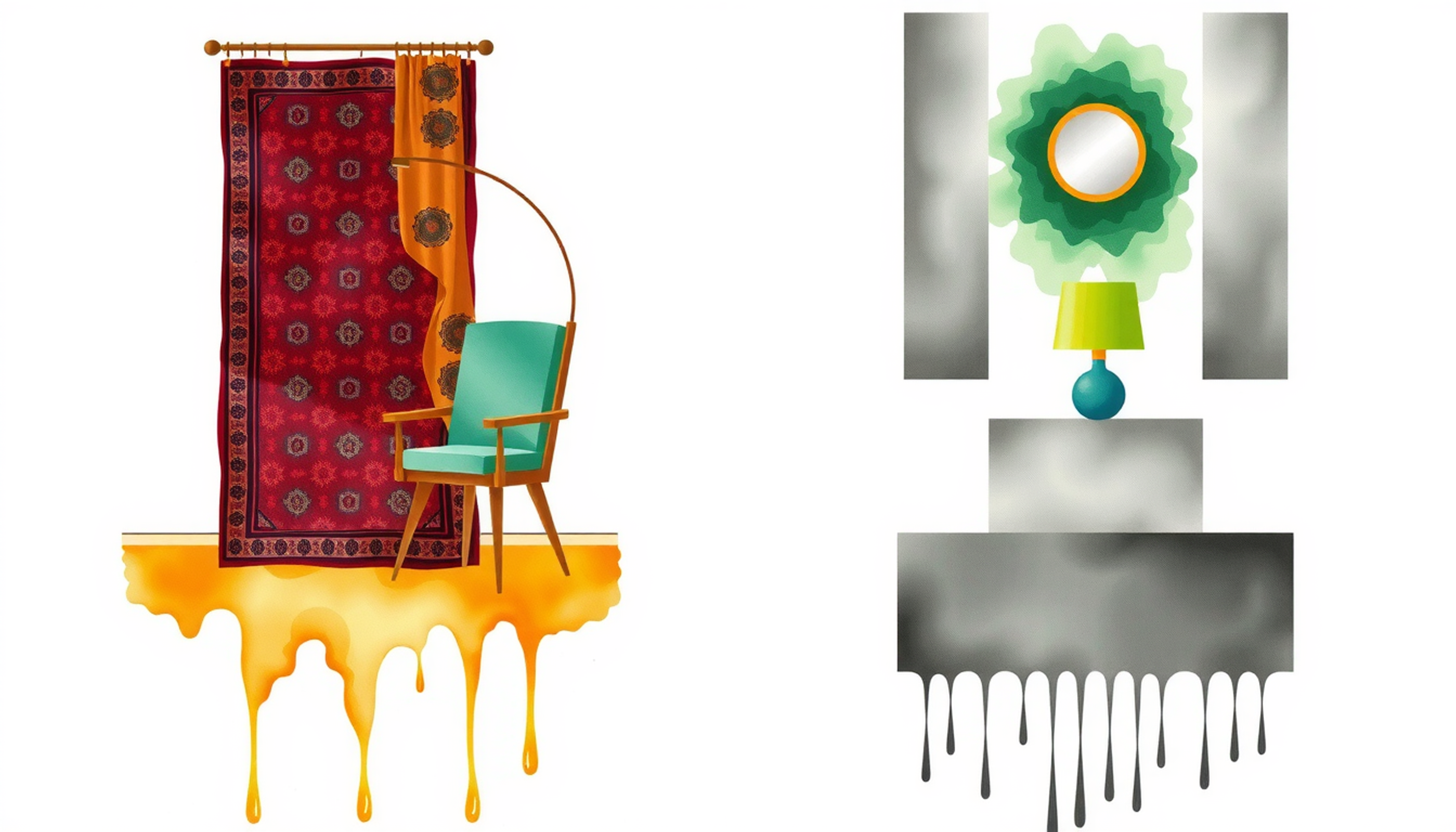
On object detail, the aphantasic group was clearly worse. Fewer objects recalled. Less colour. Where a typical imager drew a green carpet, an aphantasic participant was more likely to draw a rectangle and write what it was inside the shape. That reliance on written labels was itself a measurable difference, and it tells you something about the strategy being used.
On spatial accuracy, the two groups were equivalent. The rooms were laid out correctly. Sizes and positions were right. Whatever had gone missing, it was not the structure of the scene.
And there was a third result that nobody predicted. The aphantasic group made significantly fewer memory errors. They drew fewer objects that had not been in the rooms at all. Typical imagers, filling in their scenes from a vivid internal picture, invented more. This connects directly to what we know about how memory reconstructs rather than replays experience. A less vivid reconstruction produces a less embellished one.
The differences appeared only in the memory condition. When copying from the photograph in front of them, both groups performed the same. The bottleneck was retrieval, not perception and not drawing skill.
Read on its own, this study seems to answer the question. Aphantasia impairs object memory and spares spatial memory. Clean, quantified, large sample.
Then other laboratories ran their own experiments.
And Then the Replications Disagreed
In 2022 Pounder, who had co-authored the drawing study, published a different result in Cortex with a different team [14]. The title is blunt about what it found. Only minimal differences between individuals with congenital aphantasia and those with typical imagery on neuropsychological tasks that involve imagery.
Twenty people with aphantasia, defined by a VVIQ below 25, were matched on age and intelligence with twenty typical imagers scoring above 35. They worked through a standard automated neuropsychological battery: spatial span, a planning task, pattern recognition memory, verbal recognition memory, mental rotation.
On accuracy, almost nothing separated the groups. The only reliable difference was in how long they took. As the imagery demands of the planning and rotation tasks increased, the aphantasic group got slower. They still got the answers right.
Jianghao Liu and Paolo Bartolomeo reported something similar in Cortex in 2023 [15]. Slower responses, lower confidence ratings, but performance that held up on the tasks themselves. And in 2024 Kay, Keogh and Pearson published a mental rotation study in Consciousness and Cognition with a result that reads almost like a joke at the expense of the deficit framing: the aphantasic group was slower and more accurate, apparently because they were using a different cognitive strategy rather than a degraded version of the same one [16].
There is one further complication in the working memory literature. A 2018 case study by Christianne Jacobs, Dietrich Schwarzkopf and Juha Silvanto found that an individual with aphantasia performed worse than controls specifically on the highest-precision visual working memory trials, and showed poor insight into that performance [17]. Later group studies have generally not found working memory impairment. This is a genuinely open question, not a settled one, and it rests on a small evidence base.
Why the Disagreement Is About Method, Not Truth
It would be easy to declare a winner here. It would also be wrong. These studies are not contradicting each other on the facts. They are measuring different things and calling both of them memory.
Look at what each task actually demands.
Free-recall drawing gives you a blank page. To fill it, you have to generate the contents of a scene from nothing, in the correct detail, with no external support. That is close to the purest test of image generation anyone has designed. If image generation is the thing that is missing, this is exactly where it should show.
A recognition task gives you the answer and asks whether you have seen it before. That can be solved semantically. You do not need to reconstruct a picture. You need to consult stored knowledge, and stored knowledge is intact. The difference between these two demands maps closely onto the distinction between semantic knowledge and episodic experience, which is arguably the real fault line running through this whole literature.
Then there is what gets scored. Accuracy or speed. Almost every study that measured accuracy alone found little or no difference. Almost every study that measured latency and confidence found differences. If your outcome variable is whether the answer is right, aphantasia looks like nothing. If it is how the answer was reached and how long it took, aphantasia looks like a different route to the same destination.
Recruitment matters too. Bainbridge's participants were largely self-identified. Pounder's were selected against a strict VVIQ threshold with matched controls. Those two procedures do not necessarily capture the same population, a problem we will come back to when we get to prevalence.
And the settings differ. Large-scale online data collection with crowdsourced scoring on one side. Small, tightly controlled laboratory batteries on the other. Both approaches have well-known trade-offs and neither is obviously superior.
The synthesis that survives all of this is narrower than either camp's headline, and more useful. Aphantasia selectively affects tasks that require generating sensory detail from memory without external support. It leaves largely untouched anything that can be solved through semantic, verbal or spatial-structural routes. That is not a deficit in the amount of memory. It is a change in which retrieval strategies are available.
No one has closed this question. Anyone who tells you otherwise is reading one study.
The Memory That Thins
Something does change, though, and the clearest evidence for it comes from a different kind of memory altogether.
In 2020 Alexei Dawes, Keogh, Andrillon and Pearson published a large cognitive profile in Scientific Reports [18]. Alongside reduced imagery across several senses, the aphantasic participants reported autobiographical memories that were less vivid and less detailed. Not absent. Thinner. The events were known and datable. The experience of being back inside them was diminished.
Fraser Milton and colleagues followed in 2021 with the first systematic study combining neuropsychological testing and brain imaging across the whole imagery range: twenty-four participants with aphantasia, twenty-five with hyperphantasia, and twenty in the middle [19]. Standard memory tests showed no group differences. Autobiographical memory and imagination showed a clean gradient, with hyperphantasia above the mid-range group and aphantasia below it. Resting-state scans showed stronger connectivity between prefrontal regions and visual networks in hyperphantasia than in aphantasia.
Self-reported difficulty with face recognition was also more common in the aphantasic group, and autistic-spectrum traits scored higher. Both are associations from self-report data, not established causal links.
This thinning of autobiographical detail connects aphantasia to a separate line of research. In 2015 Daniela Palombo, Claude Alain, Hedvig Söderlund, Wayne Khuu and Brian Levine described three healthy, high-functioning adults who had never been able to relive their own past [20]. They knew what had happened to them. They could not re-experience it. Semantic memory was intact. Recognition was intact. What was missing was the first-person replay. They named it severely deficient autobiographical memory, or SDAM, and a follow-up review in 2018 set it within the wider study of individual differences in autobiographical memory [21].
The two conditions overlap heavily and they are not the same thing. Aphantasia is a trait of image generation. SDAM is a trait of autobiographical recollection. In 2018 Nicholas Watkins published an account in Cortex, both scientific and personal, of what it is to have both [22]. Some people with aphantasia have rich and detailed autobiographical memory. Some people with SDAM have perfectly ordinary imagery. The overlap is real and partial, which is exactly the sort of finding that makes clean categories difficult.
Zeman's ten-year review in Trends in Cognitive Sciences, published in 2024, gathered around fifty studies and reached a position that is deliberately restrained [1]. Imagery extremes are real, they have neural correlates, they tend to run in families, and they often extend across more than one sense. Aphantasia is variably associated with reduced autobiographical memory detail and with face recognition difficulty. Everyday functional consequences are, in his words, subtle. And a lack of imagery does not imply a lack of imagination.
That last sentence is worth sitting with, because it contradicts the way most people intuitively frame the whole subject.
Where Imagery Actually Lives in the Brain
For a long time the working assumption was that mental imagery was perception running backwards. You see something, primary visual cortex lights up. You imagine it, primary visual cortex lights up again, more weakly. Aphantasia, on that account, would be a visual cortex that fails to reactivate.
That story is no longer where the evidence points.
In 2021 Alfredo Spagna, Dounia Hajhajate, Jianghao Liu and Bartolomeo published a meta-analysis in Neuroscience and Biobehavioral Reviews covering forty-six functional imaging studies, twenty-seven of them specifically on visual imagery [23]. The consistent activation was not in early visual cortex. It was in frontoparietal control networks and in a small, well-defined region of the left fusiform gyrus corresponding to cytoarchitectonic area FG4. They named it the Fusiform Imagery Node.
The appeal of this account is that it fixes an old contradiction. Neurologists had known for decades that patients who lose mental imagery after a stroke tend to have temporal lobe damage, not occipital damage. Occipital damage causes blindness. Temporal damage takes the pictures away while leaving sight intact. The imaging literature had been pointing at the wrong place.
Joel Pearson's 2019 framework in Nature Reviews Neuroscience had already recast imagery as a top-down process whose strength varies continuously across people rather than a simple on-or-off replay [24]. The fusiform account gives that framework an anatomical anchor.
Then the evidence started converging fast.
A 7-tesla study by Liu and colleagues, published in Cortex in 2025, imaged the region at millimetre scale and found reduced functional connectivity between the Fusiform Imagery Node and prefrontal cortex in aphantasia [25]. Bartolomeo, Liu and Spagna set out the fuller argument in Neuropsychologia, describing the node as a meeting point between visual and semantic systems and proposing that the temporally correlated activity between it and left prefrontal cortex is what produces the conscious experience of an image [26]. Not the picture itself. The awareness of the picture.
The most striking result came from lesion mapping. A team including Julian Kutsche, Alexander Cohen, Michael Fox and Isaiah Kletenik conducted a systematic review of the medical literature from 1980 onwards and found twelve published cases where a brain lesion had caused loss of mental imagery [27]. They compared those twelve against 887 control lesions, using connectivity data from a thousand healthy brains.
Only five of the twelve lesions physically overlapped the Fusiform Imagery Node. All twelve were functionally connected to it. Every single one.
That is what a network finding looks like. The damage does not have to hit the node. It has to hit something wired to the node. A 2025 case study of identical twins by Emma Megla, Deepasri Prasad and Bainbridge adds another data point on the heritable and structural side, though a single twin pair is exactly the kind of evidence that needs replication before anyone builds on it [28].
There is one more finding here that refuses to sit comfortably with anything else. In 2025 Shuai Chang, Pearson, Ming Meng and colleagues published work in Current Biology reporting that visual information could be decoded from early visual cortex in aphantasic participants during imagery attempts, even though those participants reported no conscious image [29]. They called it imageless imagery.
This is contested. Liu and Bartolomeo have argued that decodable activity is not the same as imagery, because the decoded patterns lack the perception-like structure that genuine imagery produces. Whether unconscious visual representation counts as imagery at all is partly an empirical question and partly a definitional one, and the field has not resolved either half.
Counting the Uncountable
How many people have aphantasia? The honest answer is that it depends entirely on where you draw the line, and the published range reflects that rather than any real disagreement about the underlying data.
The definitive prevalence work is by Carla Dance, Alberta Ipser and Julia Simner at Sussex, published in Consciousness and Cognition in 2022 [30]. Using VVIQ scores in unselected samples, they found that 3.9 percent of people fall into a broad category of absent or dim and vague imagery. The narrower category of genuinely absent imagery came out at 0.8 percent.
Zeman's 2024 review puts extreme aphantasia at around one percent and hyperphantasia at around three percent [1]. Those figures are not in conflict with the Sussex numbers. They are answering a slightly different question.
Where the trouble really starts is that different studies use different VVIQ cutoffs. Some use 32 or below. Some use 25 or below. Some require the most extreme response on every item. Each of those choices produces a different prevalence figure and, more importantly, a different participant group. A study recruiting at 32 and a study recruiting at the floor are not studying the same people, and yet their results get compared as though they were.
Self-identification adds another layer. Some people who score in the aphantasic range on the VVIQ do not describe themselves as having aphantasia, and some who describe themselves that way score outside it. The two recruitment methods capture overlapping but distinct populations.
Andrea Blomkvist and David Marks laid this out directly in Cortex in 2023, in a paper that questioned both the measurement practice and the framing [31]. Their argument has two parts. First, arbitrary and inconsistent cutoffs are producing an artificially confused literature. Second, calling aphantasia a condition imports medical connotations that the evidence does not support, and individual difference is the better description.
The philosopher Ian Phillips pushed further in Noûs, arguing that the single vividness dimension the VVIQ measures is the wrong model altogether, and that the object and spatial components of imagery need separating before any of this can be measured properly [32].
This is contested territory and it is worth flagging as such. Zeman treats imagery extremes as real variation with neural correlates. Blomkvist, Marks and Phillips accept the variation and dispute how it is being carved up and named. Nobody in this argument thinks the phenomenon is fake. They disagree about what kind of thing it is.
What Stays Intact
Focusing on what is missing gives a distorted picture. A great deal is preserved, and the pattern of preservation is more informative than the pattern of absence.
Spatial imagery holds up. In the Keogh and Pearson study, aphantasic participants scored a mean of 19.00 on the VVIQ, far below typical, but their spatial imagery scores averaged 41.80, which was actually higher than the control mean of 36.53 [9]. Bainbridge's spatial accuracy result points the same way. Whatever the mind's eye does, it is not what handles layout, and this distinction has direct implications for how visual and spatial information get encoded in technical study material.
Semantic memory is untouched. Knowledge about the appearance of things is stored and retrievable. You know a horse has four legs, a mane and a tail. That knowledge is not a picture and never needed to be.
Recognition memory is largely preserved. Free recall is where the differences show. This is one of the cleanest patterns in the literature and it maps onto the general difference between recognising something and reconstructing it from scratch.
Perception is entirely normal. There is no visual deficit of any kind. The early stages of visual processing, including the brief sensory buffer that holds raw visual input, work exactly as they do in anyone else.
Imagery in other senses varies, and this is where a real subtyping story has emerged. In 2024 Dawes, Keogh and Pearson published a cluster analysis in Neuroscience Research covering around two thousand participants [33]. Roughly thirty percent had visual-only aphantasia, retaining imagery in at least one other modality. Roughly twenty-five percent had no imagery in any modality at all. The remainder fell into mixed and rarer patterns. Their framing, mental imagery as supramodal perception in reverse, is one of the more elegant ideas to come out of this field.
The auditory version has its own name. Rish Hinwar and Anthony Lambert introduced anauralia in Frontiers in Psychology in 2021 to describe the absence of an inner voice or inner music [34]. Whether it shows up on objective tasks the way visual aphantasia does is not yet clear, and at least one study has failed to find a difference on auditory imagery tasks. Contested, and openly so. Monzel and colleagues argued in Cortex in 2022 for consolidating the terminology under a single umbrella rather than proliferating names for each sense [35].
That prosopagnosia figure needs a note, because the number that circulates most widely is wrong. Older sources routinely quote two to two and a half percent for developmental prosopagnosia. In 2023 Joseph DeGutis and colleagues tested 3,116 adults in an unselected web sample and applied the diagnostic cutoffs actually used in the research literature [36]. The two most common criteria gave 0.93 percent and 0.45 percent. The full range across all criteria they examined ran from 0.13 to 5.42 percent, which tells you how much of the apparent prevalence of any of these traits is really a property of the measuring instrument.
Dreams, and the Line Between Voluntary and Involuntary
If you take one fact from this article to correct something you have heard elsewhere, make it this one.
Most people with aphantasia dream in pictures.
This surprises almost everyone, including people who have aphantasia and assumed their dreams must be verbal. Dawes and colleagues documented it in 2020 [18], and Zeman's review confirms that visual dreaming is usually preserved [1]. Dream imagery does tend to be reported as less controlled and less sensorily rich than in typical imagers, but it is there.
Patient MX fits the pattern from the other direction. His dreams lost their visual content and then recovered it while his waking imagery did not return [5].
The explanation is the distinction that keeps recurring through this whole subject. Aphantasia is best characterised as an absence of voluntary imagery, not an absence of all imagery. Dreams are involuntary. So are flashbacks, intrusive images and the pictures that arrive uninvited while falling asleep. Raquel Krempel and Merlin Monzel examined this directly in Consciousness and Cognition in 2024 [37], and the pattern of some involuntary imagery surviving in people with no voluntary imagery is one of the more important unresolved puzzles in the area.
It also has a practical consequence that goes well beyond dreams, since involuntary imagery is central to how emotional experience gets bound into memory.
The same voluntary and involuntary split shows up in the resting brain. The regions that generate spontaneous mental content when attention is not directed outward, described in work on the default mode network and its role in memory, remain an obvious place to look for why deliberate imagery can fail while spontaneous imagery survives. That work has not been done properly yet.

What This Means for How People Learn
This is the part of the subject where speculation runs furthest ahead of evidence, so it is worth being careful about which is which.
Start with what has actually been tested, because it produced a surprise.
Dual coding theory, one of the more durable ideas in cognitive psychology, holds that pictures are remembered better than words because they get encoded twice, once visually and once verbally. Words get one code. Pictures get two. That double encoding is supposed to explain the picture superiority effect.
If that account were right, aphantasia should abolish the effect. No visual code, no advantage.
In 2026 Muhan Yan, Brady Roberts and Bainbridge published a study in Neuropsychologia with a title that says what they found: Challenging dual-coding theory, picture superiority effects persist in aphantasia [38]. People who cannot generate mental pictures still remember pictures better than words.
This finding is frequently and incorrectly credited to the 2021 drawing study. It is not from that paper. The 2021 study examined object and spatial detail in scene recall and did not test picture superiority at all. The correct citation is the 2026 paper, and the distinction matters because the two studies point in different directions about how much imagery contributes to memory.
Reading has been tested too. Laura Speed, Lynn Eekhof and Marloes Mak published a study in Consciousness and Cognition in 2024 on the role of visual imagery in story reading, using aphantasia as a natural experiment [39]. The Wicken skin conductance work connects here as well, since the original research question came partly from people with aphantasia reporting that fiction did not move them the way it seemed to move others.
Now for what has not been tested.
The claim you will see everywhere is that people with aphantasia cannot use imagery-based mnemonics, and that the method of loci and its variants are simply unavailable to them. It is a reasonable inference. A memory palace, as classically described, involves walking through an imagined building and placing images along a route. If the images do not appear, something has to give.
But inference is not evidence, and there is very little direct experimental work on this specific question. It is entirely possible that the spatial component of the method, which is the part aphantasic participants consistently perform well on, carries more of the load than the imagery component. Nobody has run the study that would settle it. Treat any confident claim in either direction with suspicion.
What is better documented is that people with aphantasia describe compensatory strategies, and that those descriptions are consistent across studies. Verbal scaffolding. Structural and spatial encoding. Semantic elaboration. Lists. The written labels inside the drawings in Bainbridge's study are a visible trace of exactly this [13]. These are observations and self-reports rather than controlled comparisons of strategy effectiveness, and they should be read that way.
Two more claims deserve a caution rather than a repetition.
The first is that people with aphantasia gravitate toward mathematical and scientific work and away from creative fields. This comes from the 2020 Phantasia survey [7], where occupational patterns did differ between the aphantasic and hyperphantasic respondents. The problem is the sample. Everyone in it volunteered after encountering media coverage, occupations were self-reported, and there is no population base rate to compare against. It is an interesting signal from a self-selected group, not an established fact about the trait. Galton made a version of the same claim in 1880 and it did not survive scrutiny [3]. Contested, and thinly evidenced.
The second is that aphantasia protects against post-traumatic stress. Dan Cavedon-Taylor set out the theoretical case in Frontiers in Psychology in 2022, reasoning that if intrusive imagery maintains certain disorders, then an absence of imagery might be protective [40]. The Wicken result gives that reasoning a plausible mechanism. What does not exist is clinical outcome data showing lower rates of the disorder in aphantasic populations. The hypothesis is live. The conclusion is not established.
There is a better-supported point buried in the same area. Many of the standard psychological treatments for trauma and anxiety assume that the person can generate mental images on demand. Imaginal exposure. Imagery rescripting. A 2024 study in Collabra by Bridget Mawtus and colleagues documented the practical consequences of that assumption for people who cannot do it [41]. That is a real and actionable finding, and it is considerably firmer than the protection hypothesis it gets bundled with.
Condition or Difference
Ten years after the word was coined, the field has not settled what kind of thing it named.
One position, broadly Zeman's, is that imagery vividness sits on a continuum with meaningful extremes at both ends, that those extremes have measurable neural and physiological correlates, and that they deserve names [1]. On this account, aphantasia is a real variation with real consequences, most of them subtle.
The other position, argued by Blomkvist and Marks, is that calling it a condition smuggles in a medical frame that the evidence does not carry, and that a decade of inconsistent measurement has produced a literature where studies cannot be properly compared [31]. Phillips adds that the underlying construct is probably not one dimension at all [32].
These positions are less far apart than they sound. Nobody disputes that some people do not generate voluntary mental images. The argument is about whether that fact is best described as a variation, a condition, or a cluster of partly independent traits that happen to correlate.
There is a version of this that is not academic hair-splitting. Acquired loss of imagery after a stroke or brain injury sits in genuinely different territory from a lifelong trait, and the lesion mapping work treats it as such [27]. Lumping the two together under one word has caused confusion in both directions.
Conclusion
The most useful thing aphantasia has done for memory science is act as a natural dissociation. It separates things that normally travel together, and the separation reveals which of them were doing the work.
Take mental pictures out of a mind and semantic knowledge stays. Recognition stays. Spatial layout stays. Perception is untouched. Dreams, mostly, keep arriving. Picture superiority persists, which dual coding theory did not predict. Autobiographical memory thins without disappearing, and object detail in free recall drops while spatial accuracy does not.
That is a very specific pattern, and it does not look like damage. It looks like a different configuration.
The open questions are real ones. Whether the object memory difference is reliable or task-dependent is not resolved, and Bainbridge and Pounder are both still right about their own data. Whether decodable activity without conscious experience counts as imagery is unsettled. Whether the trait deserves a clinical frame is a live argument between serious people. Whether imagery-based memory techniques are genuinely unavailable is close to untested, despite being asserted constantly.
What has changed is that these are now empirical questions rather than philosophical ones. In 2015 there was a three-page paper and twenty-one letters. Now there is pupillometry, lesion network mapping, 7-tesla imaging and prevalence data from unselected population samples. That is fast, by the standards of any field.
And the finding that should be hardest to forget is the smallest one. In the pupil study, when aphantasic participants were asked to imagine four things instead of one, their pupils widened [12]. The effort was there. The load was there. The machinery was running. Only the picture never came.
Memory, it turns out, was never mainly about pictures.
Frequently Asked Questions
Does aphantasia mean someone has a bad memory?
No. Research consistently shows intact semantic memory, preserved recognition, and spatial accuracy equal to typical imagers. What differs is the richness of autobiographical recall and performance on tasks requiring people to generate visual detail from scratch. Memory capacity is not reduced. The retrieval strategy differs.
Can people with aphantasia see images in their dreams?
Most can. Studies by Dawes and colleagues and Zeman's ten-year review both report that visual dreaming is usually preserved, though dream imagery tends to be described as less controlled and less sensorily rich. Aphantasia is best understood as an absence of voluntary imagery rather than all imagery.
How common is aphantasia in the general population?
Estimates range from roughly 0.8 percent to 3.9 percent depending entirely on the definition used. Sussex researchers found 3.9 percent for absent or dim imagery and 0.8 percent for completely absent imagery. Zeman's review places extreme aphantasia near one percent and hyperphantasia near three percent.
How do researchers prove aphantasia is real and not just poor self-reporting?
Three independent physiological measures. Binocular rivalry priming sits at chance. Skin conductance stays flat during frightening text but responds normally to frightening images. Pupils fail to track imagined brightness while still dilating with cognitive load, showing that mental effort occurs even when no image is produced.
Which brain region is involved in aphantasia?
Current evidence points to the Fusiform Imagery Node in the left fusiform gyrus and its connection to prefrontal cortex, not to early visual cortex as once assumed. A lesion mapping study found that all twelve documented cases of acquired aphantasia involved damage functionally connected to this node.