

INTRODUCTION
For most of the twentieth century, neuroscience believed a lie. The lie was simple: after childhood, the adult brain is fixed. No new neurons are built. No new connections are formed. What you have is what you will always have. This idea shaped how we designed education systems, how we treated stroke patients, and how we thought about aging. And it was wrong. The human brain contains roughly 86 billion neurons [1] and somewhere between 100 and 500 trillion synaptic connections. And it rewires itself constantly. From the day you are born until the day you die. This ability is called neuroplasticity, and understanding it changes everything. How we think about learning. How we treat brain injuries. What we tell a sixty-year-old who wants to learn a new language. But here is the part that rarely makes headlines: plasticity is not always your friend. The same mechanism that lets your brain recover from a stroke is the one that builds chronic pain, deepens addiction, and locks in trauma. Plasticity is a sculptor. It can build or destroy. It does not care which.
The Biggest Mistake in the History of Brain Science
This is a story about a wrong idea that lasted over a hundred years.
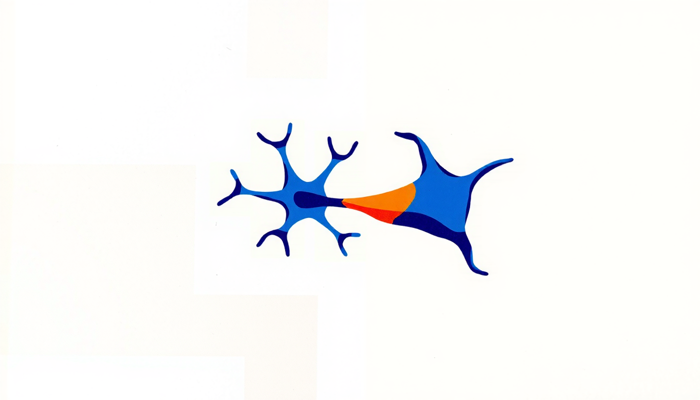
Santiago Ramón y Cajal won the Nobel Prize in 1906 for proving that the nervous system is made of individual cells — neurons — not a continuous web. He is the father of modern neuroscience. In his 1928 book, he wrote a sentence that became gospel: once development ends, the sources of growth and regeneration of axons and dendrites — the branching arms that neurons use to communicate — dry up irrevocably [2]. That one sentence froze the field for decades.
But here is something almost nobody mentions. Cajal himself said the opposite thirty-four years earlier. In his 1894 Croonian Lecture at the Royal Society of London, he stated that neurons in adults have the ability to grow and that new connections could explain learning [2]. He even used the phrase "neuronal plasticity" in 1904 [3]. The dogma that halted a century of progress was a selective reading of a man who had argued both sides.
Even before Cajal, someone got it right. William James — the philosopher-psychologist at Harvard — wrote in The Principles of Psychology in 1890 that organic matter, especially nervous tissue, seems to possess an extraordinary degree of plasticity [4]. He defined plasticity as having a structure weak enough to yield to an influence, but strong enough not to yield all at once. That was 1890. The field took another century to reach where James had been standing.
So what happened? Why did the wrong idea win? Partly because the right tools did not exist. You cannot see synapses forming without electron microscopes. You cannot watch cortical maps reorganize without microelectrode arrays. And partly because the wrong idea was more comfortable. If the brain is fixed, then neurology becomes simple: damage is permanent, decline is inevitable, and there is not much to be done.
The cracks started in the 1960s. Joseph Altman at MIT injected radioactive thymidine — a molecule that gets incorporated into new DNA during cell division — into adult rats and found labeled neurons in the hippocampus. The hippocampus is a seahorse-shaped structure deep in the brain that serves as the memory factory [5]. This meant the adult brain was making new neurons. The field ignored him. His finding was so contrary to the dogma that it was simply dismissed.
Then Marian Diamond at UC Berkeley ran a beautiful experiment. She placed rats in two environments: one enriched with toys, tunnels, and other rats, and one impoverished with nothing but food and water. After several weeks, she measured their brains. The enriched rats had cerebral cortex seven to ten percent heavier, roughly twenty percent more synaptic connections, and even synapses fifty percent larger than those of the impoverished rats [6]. This was the first solid proof that experience physically changes the brain. Diamond published in Science. 1964. It was replicated multiple times. And still the dogma held.
The Rebels Who Proved the Brain Rewires
The final assault on the fixed-brain dogma came from two unlikely sources: a man who helped the blind see through their skin, and a man whose monkey experiments sparked the animal rights movement.
Paul Bach-y-Rita was a neuroscientist at the Smith-Kettlewell Institute in San Francisco. In 1969, he built something that sounds like science fiction. He took a dental chair, embedded 400 vibrating metal plates in the backrest, and connected them to a camera. When a blind person sat in the chair, the camera image was translated into vibration patterns on their back. With practice, blind subjects could recognize faces, track moving objects, and even catch a ball rolling across a table [7].
Think about what this means. The visual cortex — the part of the brain whose job is processing sight — was receiving information through the skin. Not through the eyes. The brain was reassigning an entire sensory region to process a completely different kind of input. Bach-y-Rita published in Nature. Reviewers thought he was crazy. He spent years defending the work. But he had demonstrated something profound: the adult brain does not care where information comes from. What matters is what the information means. And it will rewire itself to process it.
Michael Merzenich at UC San Francisco delivered the most technically rigorous evidence. In 1983 and 1984, he used dense microelectrode arrays — dozens of tiny wires inserted into the brain — to map the somatosensory cortex of adult owl monkeys. The somatosensory cortex is the strip of brain that processes touch. Each finger has its own small patch of cortex, like a tiny map. Merzenich amputated a single digit and then re-mapped the cortex weeks later. The patch belonging to the missing finger was not silent. It had been invaded by the patches of adjacent fingers [8], [9].
This was not healing. This was not repair. This was reorganization. The adult cortex had redrawn its own map based on what information was available. Merzenich's work, independently confirmed by Jon Kaas and others, made the case undeniable: the adult brain is not hardwired. It is dynamically maintained. Change the input, and the map changes.
The implication is enormous. If cortical maps can reorganize after injury, they can also reorganize during learning. Every time you practice a skill, memorize a fact, or navigate a new city, your brain is physically rewriting itself. The question stopped being whether the brain can change. It became: how does it change, how much, and can we control it?
The Rule That Explains How You Remember Anything
In 1949, a Canadian psychologist named Donald Hebb published a book called The Organization of Behavior. It contained one idea that changed neuroscience forever. Hebb proposed that when neuron A repeatedly helps fire neuron B, the connection between them strengthens [10]. The popular summary is five words: neurons that fire together, wire together.
This sounds simple. It is not.
Hebb was saying something radical for 1949. He was saying that memories are not stored in individual neurons like files in a cabinet. They are stored in the pattern of connections between neurons — what he called "cell assemblies." When you remember your first day of school, no single neuron holds that memory. Instead, a specific constellation of neurons fires together, and the strength of their connections is the memory itself.
But Hebb was making a theoretical prediction. He had no way to prove it. The proof came twenty-four years later, in a laboratory in Oslo, and it came by accident.
In 1973, Timothy Bliss from London and Terje Lømo from Norway were working in Per Andersen's lab, studying a brain structure called the hippocampus — the seahorse-shaped region deep in the temporal lobe that serves as the brain's memory factory. They were studying ordinary synaptic transmission in anesthetized rabbits when Lømo delivered a burst of high-frequency electrical stimulation to a neural pathway called the perforant path. Something unexpected happened. After the stimulation stopped, the synapses did not go back to normal. They stayed stronger. For hours. In some cases, more than ten hours [11].
They had discovered long-term potentiation, or LTP. It was the first physical mechanism that could explain how memories form at the level of individual synapses. Use a connection, and it gets stronger. The effect lasted far longer than the stimulation itself. Lømo later revealed that he had first observed the phenomenon in 1966 during his thesis work but did not understand what he was seeing [12]. One of the most important discoveries in neuroscience happened twice, and the first time, nobody noticed.
But strengthening is only half the equation. The brain also needs to weaken connections that are no longer useful. In 1982, Masao Ito at the University of Tokyo discovered the mirror image of LTP: long-term depression, or LTD. Specific patterns of low-frequency stimulation cause synapses to become weaker and stay weaker [13]. LTP and LTD together give the brain a volume knob for every single synapse. Turn connections up. Turn connections down. This is how you learn. This is also how you forget.
The molecular key to both processes is a protein called the NMDA receptor. Think of it as a coincidence detector. It sits on the surface of neurons and has a peculiar property: it only opens when two things happen simultaneously — a chemical signal arrives from the sending neuron, AND the receiving neuron is already active [14]. Both conditions. At the same time. If only one occurs, nothing changes. This is the molecular implementation of Hebb's rule: the NMDA receptor only strengthens a connection when the two neurons are firing together. Graham Collingridge proved this in 1983 by showing that blocking NMDA receptors prevents LTP without affecting normal synaptic transmission [14].
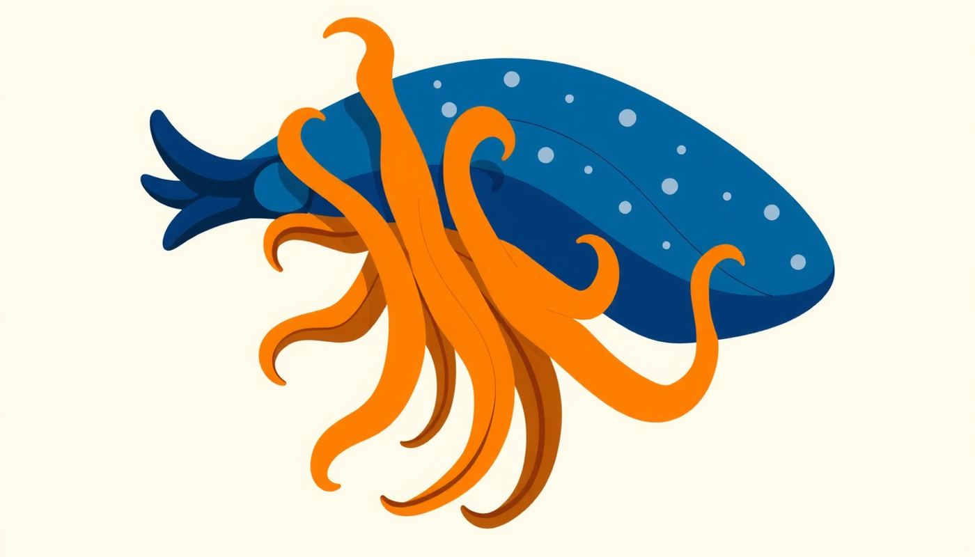
Now here is where the story gets its Nobel Prize. Eric Kandel at Columbia University wanted to understand memory at the molecular level. But the human brain is hopelessly complex. So he chose the simplest nervous system he could find: a sea slug called Aplysia californica. It has only about twenty thousand neurons, and they are large enough to see with the naked eye. By studying how Aplysia learns to retract its gill from a threatening touch, Kandel traced the entire molecular pathway of memory formation.
What he found was beautiful in its simplicity. Short-term memory — the kind lasting minutes to hours — involves only chemical changes at existing synapses. An enzyme called protein kinase A modifies potassium channels, causing more neurotransmitter to be released. No new structures are built. But long-term memory — the kind lasting days, weeks, or a lifetime — requires something fundamentally different. The enzyme travels to the cell nucleus, activates a gene-switching protein called CREB, which turns on genes that build new proteins, which construct entirely new synaptic connections [15]
In other words: short-term memory is a software update. Long-term memory is a hardware upgrade.
Kandel won the Nobel Prize in Physiology or Medicine in 2000. And his finding established a principle that runs through everything in this article: lasting changes in the brain require structural changes. Not just different chemistry. Different architecture. This principle explains why spaced repetition works: each time you review information, the relevant synapses are strengthened and new physical structures are built. Without repetition, connections stay weak and the memory fades.
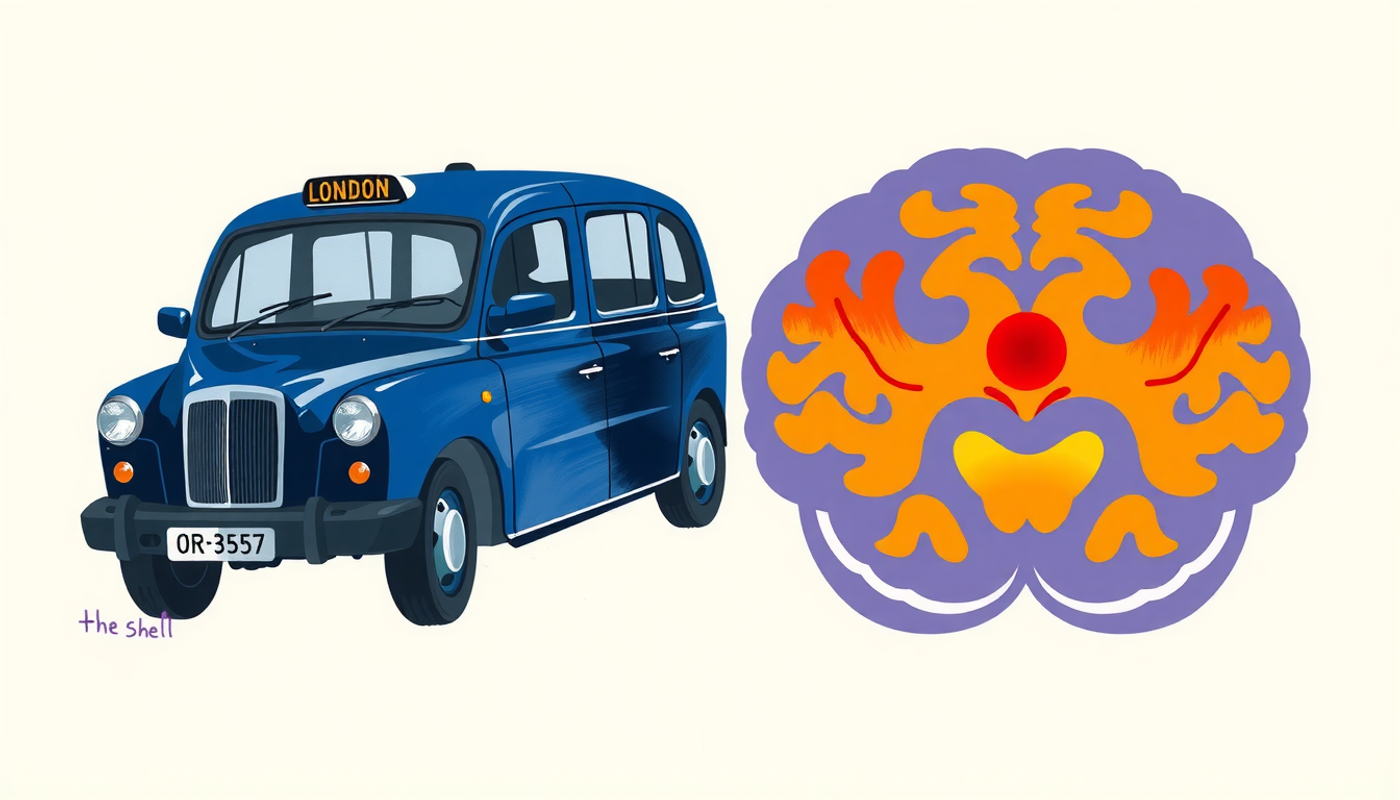
The Taxi Drivers Whose Brains Got Bigger
Perhaps the most iconic evidence for human neuroplasticity came from the streets of London. Not from a lab.
Eleanor Maguire at University College London scanned the brains of sixteen male London taxi drivers using MRI in 2000 and compared them with fifty controls. London taxi drivers must pass an exam called "the Knowledge" that requires memorizing over twenty-five thousand streets and thousands of landmarks within a six-mile radius of central London. It takes three to four years. The MRI result was striking: the posterior hippocampus of taxi drivers was significantly larger, and its size correlated positively with years of driving experience [16].
But one big question remained: maybe these people had different brains to begin with, and that is why they became taxi drivers? Maguire answered this in 2006 by comparing taxi drivers with bus drivers. Both groups drove in London for hours, but only taxi drivers had to navigate. Bus drivers showed none of the hippocampal changes [17]. Spatial knowledge, not driving itself, was changing the brain.
The decisive blow came in 2011. Maguire scanned seventy-nine taxi trainees and thirty-one controls before training, then again three to four years later. At baseline, no structural differences existed between any of the groups. After training, only the thirty-nine who qualified showed increased gray matter in the posterior hippocampus. The forty who failed showed nothing [18].
This proved the changes were caused by learning, not genetics. But there was another interesting finding: the qualified taxi drivers performed worse on a visual memory test. The brain had made the posterior hippocampus bigger by making the anterior hippocampus smaller. Expertise has a cognitive cost. The brain simply shuffles resources around.
Does the Adult Brain Make New Neurons? The Most Heated Debate in Neuroscience
Here is where things get really heated.
Until the late twentieth century, everyone thought the adult brain could not make new neurons. Then in 1998, Fred "Rusty" Gage at the Salk Institute used BrdU — a molecule that gets incorporated into dividing DNA — to find new neurons in the hippocampus of five deceased cancer patients. About twenty-two percent of the labeled cells were neurons [19]. The adult human brain was making new neurons. A revolution.
Then everything blew up. In March 2018, the team of Shawn Sorrells and Arturo Alvarez-Buylla at UCSF published a paper in Nature. They examined fifty-nine human brain tissue samples from prenatal to age seventy-seven. Their conclusion was jarring: young neurons dropped sharply in the first year of life and were virtually undetectable in adults [20].
Four weeks later — just four weeks — the team of Maura Boldrini at Columbia published a paper in Cell Stem Cell. They examined twenty-eight healthy brains from ages fourteen to seventy-nine and found the exact opposite: similar numbers of neural progenitors and thousands of immature neurons at all ages [21].
Both teams used human tissue. Both were credible. The field exploded.
The key to solving the puzzle came from Madrid. María Llorens-Martín showed that storing brain tissue in formalin for more than six months completely destroys the DCX marker — a protein used to identify young neurons. Even in infant tissue where neurogenesis is beyond dispute. None of nine commercially available anti-DCX antibodies worked after prolonged fixation. In her own carefully controlled samples, she found thousands of immature neurons into the ninth decade of life [22].
And in 2025, a major breakthrough arrived. Jonas Frisén's team at the Karolinska Institute in Sweden used an entirely different technology: single-nucleus RNA sequencing. This method does not depend on the problematic DCX antibodies at all — it directly reads the active genes in each cell. In thirty-five individuals from age zero to seventy-eight, they identified actively dividing neural progenitor cells in the adult hippocampus [23].
Where does this leave us? The weight of evidence now tilts heavily toward adult neurogenesis continuing in humans. A 2018 consensus statement signed by eighteen prominent researchers supports this conclusion [24]. The Alvarez-Buylla team still maintains its skepticism. Science sometimes works this way. Evidence accumulates, but consensus arrives slowly.
When Learning Physically Rewires the Brain
London taxi drivers are not the only example. The evidence comes from everywhere.
Professional musicians have a larger corpus callosum — the bridge of nerve fibers connecting the two brain hemispheres. But this effect was only found in those who started training before age seven [25]. Musicians also show increased gray matter in motor, auditory, and spatial regions [26].
Perhaps the cleanest demonstration came from Bogdan Draganski in 2004. Twenty-four people who had never juggled were split into two groups. Twelve learned to juggle three balls for three months. Twelve did nothing. After three months, the jugglers' brains showed significant gray matter increases in two regions: area hMT/V5, which processes visual motion, and the left posterior intraparietal sulcus, which manages hand-eye coordination. After three months without practice, the changes shrank back toward baseline [27].
Think about that. Three months of learning a simple skill created measurable changes on MRI. And when practice stopped, the brain started reverting. Plasticity runs both ways: "use it or lose it" is not just a saying. It is biology.
Learning a second language changes the brain too. Bilinguals have greater gray matter density in the left inferior parietal cortex, and the degree of change correlates with proficiency and age of acquisition [28]. Even German medical students preparing for preliminary exams showed gray matter increases in the posterior hippocampus and parietal cortex — and the hippocampal changes continued growing three months after the exam [29]. As if the brain, once freed from exam stress, had the opportunity to complete changes that began under pressure. This aligns with what we know about the effects of stress on memory: chronic cortisol suppresses hippocampal plasticity, but after the stressor is removed, the brain can recover.
The message is clear. Every form of deep learning — language, music, navigation, even juggling — physically changes the brain. The changes are small (one to five percent by volume) but real and measurable.
Windows That Close — And People Who Reopen Them
A kitten whose eye is sewn shut will lose vision in that eye forever. An adult cat under the same conditions? No damage at all. This difference introduced the concept of the "critical period."
David Hubel and Torsten Wiesel at Harvard — Nobel laureates in 1981 — showed that during the first weeks of a kitten's life, visual cortex neurons must receive input from both eyes or the ocular dominance columns permanently reshape. Cells in the lateral geniculate nucleus — the visual relay station between the eye and cortex — shrank by about forty percent in the deprived eye. But the same deprivation in an adult cat had no effect [30]. A time window existed. And then it closed.
The same logic applies to language. An experiment with forty-six Korean and Chinese immigrants to the US found a strong linear decline in English proficiency with age of arrival up to puberty — after puberty, performance became irregular and weaker [31].
But can these windows be reopened? Takao Hensch at Harvard identified the molecular "brakes" on plasticity: perineuronal nets — mesh-like protein structures that wrap around mature neurons and prevent change. Remove these brakes or inhibit their enzymes, and adult visual cortex becomes plastic again [32]. In 2013, a double-blind trial showed that valproic acid — an anti-seizure drug — helped adult men learn absolute pitch, a skill previously thought learnable only in childhood [33].
And in 2023, a landmark paper in Nature showed that psychedelics — psilocybin, LSD, ketamine, MDMA, and ibogaine — all reopen the social reward learning critical period in adult mice. The duration of reopening was proportional to the subjective duration of each drug's effects in humans. The mechanism involved extracellular matrix remodeling and changes in expression of roughly sixty-five genes [34].
This means learning windows are not necessarily closed forever. It may be possible to pharmacologically reopen them. The field is still early, but the implications for stroke recovery, developmental disorders, and even adult language learning could be immense.
Exercise: The Most Powerful Plasticity Drug You Can Get Without a Prescription
Of everything that enhances brain plasticity, exercise has the strongest evidence.
The molecular key is a protein called BDNF — brain-derived neurotrophic factor. Think of it as fertilizer for neurons. BDNF facilitates long-term synaptic potentiation, stimulates dendritic growth, and supports new neurons. Aerobic exercise drives BDNF levels up [35].
But this is not just molecular talk. The human evidence is strong. Kirk Erickson's randomized controlled trial with 120 older adults (average age sixty-seven) showed that one year of aerobic walking — forty minutes a day, three times a week — increased left hippocampal volume by 2.12 percent and right by 1.97 percent. The control group doing stretching exercises declined by 1.4 percent. That two percent increase effectively reversed one to two years of age-related brain shrinkage. And the volume increase correlated with higher serum BDNF [36].
A meta-analysis of twenty-nine studies found that even a single exercise session raises BDNF with a moderate effect size of 0.46 [37]. And another meta-analysis of fourteen studies with 737 participants confirmed significant positive effects of aerobic exercise on left hippocampal volume [38].
Scientific honesty requires a note: a more conservative 2024 meta-analysis of eight randomized trials in healthy older adults found the effect on hippocampal volume did not quite reach statistical significance. Exercise is undoubtedly beneficial, but the structural effect on the brain may be more modest than early media headlines suggested. That is what honest science looks like.
When Plasticity Becomes the Enemy
So far, neuroplasticity might sound like a superpower. But the same mechanism that allows your brain to learn and recover can work against you.
V.S. Ramachandran at UC San Diego discovered that after arm amputation, the hand area in the brain gets invaded by adjacent regions — especially the face. Touching specific points on a patient's face produced sensations in phantom fingers [39]. Herta Flor showed the extent of cortical reorganization correlated directly with phantom limb pain intensity [40]. The brain was rewiring itself, but the result was pain, not recovery.
Chronic pain itself is a form of maladaptive plasticity. Central sensitization — increased excitability of pain neurons in the spinal cord — uses the same LTP-like mechanisms. Pain synapses get stronger. Pain thresholds drop. The pain system literally learns to produce more pain [41]. The good news: gray matter changes in chronic pain patients are reversible with effective treatment.
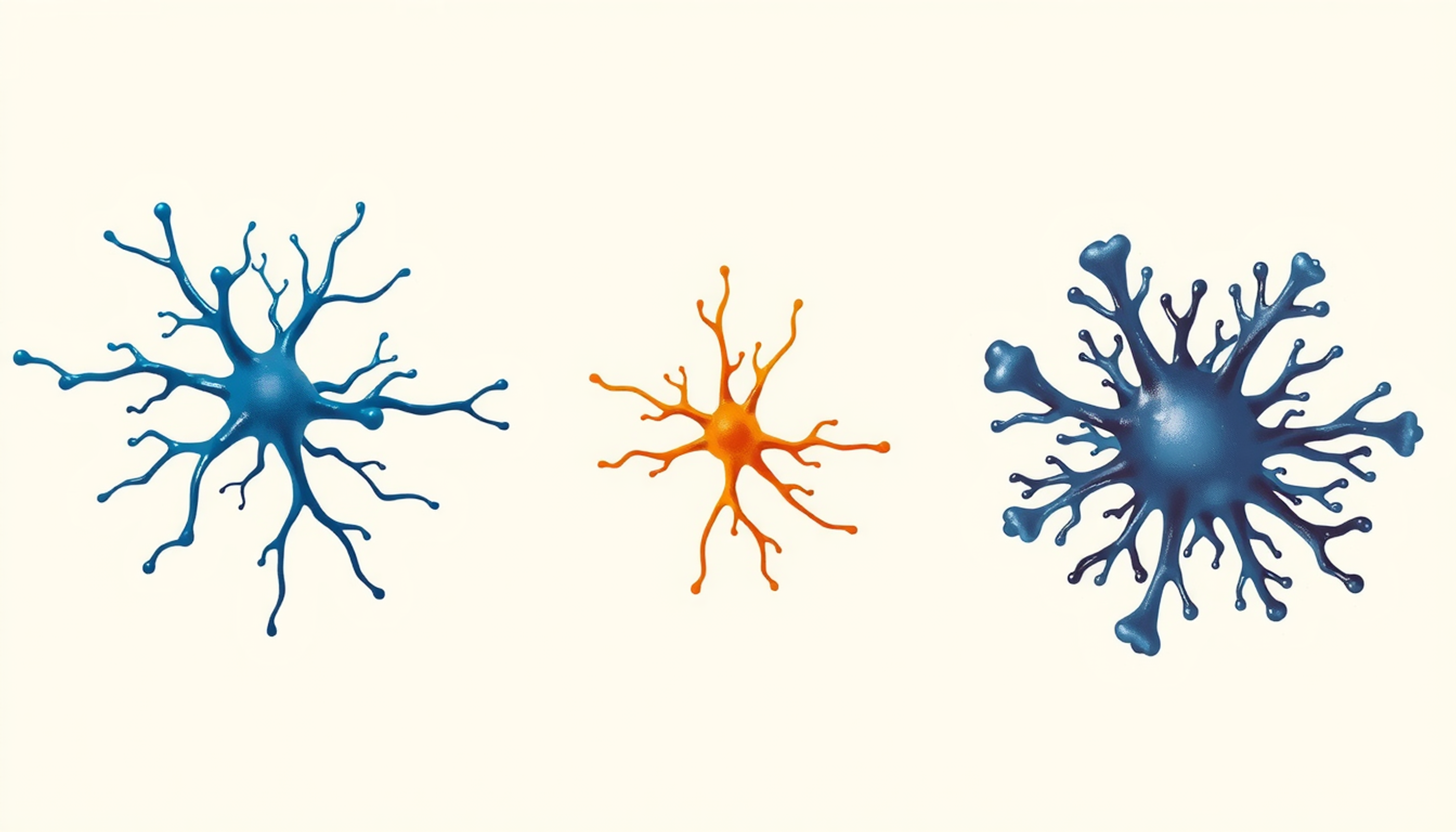
Addiction is the same story. Drugs of abuse hijack plasticity mechanisms. Cocaine increases the number of dendritic spines — the tiny protrusions on neuronal branches where synapses form — in the brain's reward center. A protein called ΔFosB persists for weeks after the last dose, keeping reward circuits sensitized [42]. Addiction is extraordinarily powerful and extraordinarily destructive learning.
Even tinnitus — that constant ringing experienced by ten to fifteen percent of adults — is an example of misguided reorganization. After hearing damage, the auditory map in the brain restructures, and frequencies that no longer receive input get taken over by neighboring frequencies. Spontaneous firing rates in reorganized areas increase by up to forty percent [43]. The result: a sound that has no external source, but the brain hears it anyway.
Neuroplasticity has no morals. It is just a tool. And like any tool, what it produces depends on how it is used.
Sleep: The Price the Brain Pays for Plasticity
Wakefulness learns. Sleep consolidates. Without one, the other is useless.
Giulio Tononi and Chiara Cirelli at the University of Wisconsin-Madison proposed the "synaptic homeostasis hypothesis": during wakefulness, learning causes net synaptic strengthening. During deep sleep — slow-wave sleep — synapses are globally downscaled to restore energy balance, signal-to-noise ratios, and learning capacity. Their memorable formulation: "Sleep is the price the brain pays for plasticity" [44].
The ultrastructural evidence came in 2017. Tononi's team measured 6,920 synapses in mouse cortex using electron microscopy. The axon-spine interface was about eighteen percent smaller after sleep compared to wakefulness. But the downscaling was selective: about eighty percent of weaker synapses shrank, while the strongest twenty percent were spared [45]. Sleep clears the noise but keeps the important signals.
Memory consolidation during sleep works through the coordination of three brain oscillations: slow oscillations from the prefrontal cortex, sleep spindles from the thalamus, and hippocampal sharp-wave ripples. When these three synchronize, memories are transferred from the hippocampus to the cortex. Selectively suppressing hippocampal ripples during post-learning sleep impaired spatial memory in rats [46].
This is where the connection to stress and memory becomes important. Chronic stress keeps cortisol elevated, elevated cortisol disrupts deep sleep, and without deep sleep, memory consolidation does not happen. A vicious cycle that starts with stress and ends with an inability to learn.
And a practical takeaway: any learning method based on spaced repetition — reviewing information at increasing intervals — works precisely because it gives sleep the opportunity to perform synaptic consolidation between review sessions. Without that spacing, sleep cannot do its job.
Stress and Plasticity: Builder or Destroyer?
Bruce McEwen at Rockefeller University spent five decades mapping how stress reshapes brain architecture. His most striking finding: twenty-one days of chronic stress shortened the dendrites of hippocampal pyramidal neurons by twenty to thirty percent. But the same stress had the opposite effect in the amygdala — the brain region that processes threat — where dendrites grew and synapses multiplied [47].
This means chronic stress does not destroy the brain. It reorganizes it. Resources are shifted away from memory and flexible thinking (hippocampus and prefrontal cortex) and toward threat detection (amygdala). When threats are real and temporary, this is adaptive. When they are chronic, it is devastating.
But McEwen showed something else that matters: after the twenty-one days of stress ended, if the animals were given ten to twenty-one days of rest, hippocampal dendrites regrew [48]. The damage was reversible. Exercise and enriched environments accelerated recovery. Chronic stress is not a life sentence. The brain has the capacity to heal. You just have to give it the opportunity.
Myths That Need to Die
Neuroplasticity is real. But the self-help industry and marketing have weaponized it. Time to bury some myths.
"You only use ten percent of your brain." This probably comes from William James who wrote in 1908 that we use only a small part of our possible mental resources. His statement was about potential, not brain tissue. Brain imaging shows virtually all regions are active within twenty-four hours. The brain consumes twenty percent of the body's energy while comprising only two percent of its weight. Evolution would never maintain an organ this expensive if ninety percent of it were idle.
"Brain training games make you smarter." On October 20, 2014, more than seventy leading cognitive scientists from the Stanford Center on Longevity and the Max Planck Institute released a joint statement: the scientific literature does not support claims that software-based brain games improve general cognitive performance in everyday life. One hundred and thirty-three people wrote a counter-letter — but some signatories had financial ties to the brain training industry.
In January 2016, the US Federal Trade Commission fined Lumosity two million dollars for deceptive claims about preventing Alzheimer's and memory loss. And the largest brain training study to date — 11,430 participants in collaboration with the BBC — found no evidence of transfer to untrained tasks, even when those tasks were cognitively similar [49]. The most comprehensive review concluded: extensive evidence for improvement on practiced tasks, less evidence for closely related tasks, and virtually no evidence for everyday cognitive improvement [50].
So what actually works? Aerobic exercise has the strongest evidence. Learning complex new skills — languages, musical instruments, navigation — produces measurable structural changes. Adequate sleep. Social relationships. And among cognitive interventions, speed-of-processing training shows the most promising transfer effects.
CONCLUSION
The story told in this article is more complex than "the brain can change."
Yes, the adult brain is not hardwired. That is now beyond dispute. From Diamond's lab in 1964 to Maguire's taxi driver scans to 2025 imaging finding new neurons in the hippocampus of eighty-year-olds, the evidence is undeniable. Every time you learn something, your brain physically changes. Synapses strengthen, dendrites grow, and in some regions, new neurons may be born.
But plasticity is not a superpower without cost. The same mechanisms build chronic pain, deepen addiction, and produce tinnitus. Expertise in one area comes at a cognitive cost in others — London taxi drivers had smaller anterior hippocampi. Chronic stress reorganizes the brain in ways that weaken memory and amplify anxiety.
And a multibillion-dollar industry profits from exaggerated promises about plasticity. Brain training games and "brain health" supplements make claims that far exceed the evidence. Seventy Stanford scientists made this clear.
What the evidence actually shows is simpler and yet more profound: aerobic exercise, learning complex new skills, adequate sleep, and social connection — these are the real pillars of brain maintenance. And perhaps the most important message is that the brain can heal. Hippocampi that shrank under stress regrow with rest and exercise. This is not a miracle and it is not a slogan. It is biology.
Frequently Asked Questions
Does neuroplasticity continue in old age?
Yes. Research confirms the brain remains plastic throughout the entire lifespan, though the speed and extent of changes decrease with age. Studies of London taxi drivers, elderly jugglers, and exercise interventions in adults over sixty-five have all demonstrated measurable structural brain changes.
Is stress-related brain damage reversible?
Animal studies show twenty-one days of chronic stress shrinks hippocampal dendrites by twenty to thirty percent, but ten to twenty-one days of rest restores them. In humans, Cushing's syndrome patients showed hippocampal regrowth after treatment. Exercise and enriched environments accelerate recovery.
Do brain training games actually work?
The largest study (11,430 participants with the BBC) found no evidence of transfer to untrained tasks. Over seventy cognitive scientists from Stanford and Max Planck issued a 2014 statement rejecting claims of general cognitive improvement. Aerobic exercise has far stronger evidence for brain health.
Has adult neurogenesis been proven in humans?
The weight of evidence now supports continued neurogenesis in the adult hippocampus. Independent methods including BrdU labeling, controlled-fixation immunohistochemistry, and single-nucleus RNA sequencing have all found new neurons. But the exact rate and functional significance remain debated.
What is the best evidence-based way to boost neuroplasticity?
Three interventions have the strongest evidence: regular aerobic exercise that increases hippocampal volume and BDNF levels, learning complex new skills such as languages or musical instruments, and sufficient quality sleep essential for synaptic consolidation. Social engagement is consistently protective as well.


