

Introduction
Every night something strange happens. You put your head on the pillow. Your eyes close. And apparently nothing happens. But if electrodes were attached to your skull, you would see a completely different picture. Your brain has not only not shut down — at certain moments it is more active than during wakefulness [1]. And it is not doing just one thing. It is simultaneously sorting the day's memories, rewriting emotions, washing away waste proteins, listening to outside sounds, and in some of its regions even staying awake while the rest has gone into deep sleep. Until just twenty years ago, most scientists considered sleep a single, uniform state. The brain was either awake or asleep. Black or white. But today the picture has completely changed. New research using intracranial recordings, brain imaging, and optogenetics has shown that sleep is more like a mosaic [2]. Different brain regions can simultaneously be in different states. Some in deep sleep. Some in light sleep. Some practically awake. And each doing a different job. This article tells the story of the discovery of this mosaic. The story of scientists who realized the sleeping brain is not one mind at rest. It is a factory with multiple production lines running around the clock.
The Night Neuroscience Woke Up
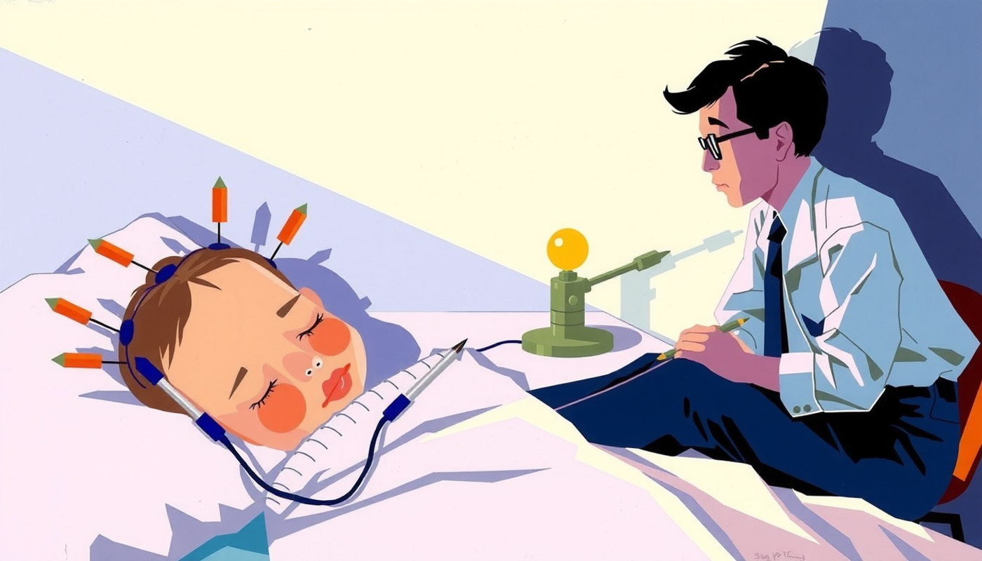
The history of sleep science has a clear starting point. Fall 1952. Eugene Aserinsky, a twenty-nine-year-old doctoral student at the University of Chicago, was recording the sleep of his eight-year-old son Armond with an electroencephalography machine. His advisor was Nathaniel Kleitman, the man who would later be called the father of sleep research. Aserinsky expected the machine's pens to draw calm, uniform lines. But in the middle of the night, the pens went wild. Fast, irregular lines were drawn on the paper, as if the child had woken up and was looking around. But Armond was asleep [3].
Aserinsky first thought the machine was broken. He checked the wires. Recorded again. Same result. The child's eyes were moving rapidly under closed eyelids. His brain showed a pattern resembling wakefulness. But his body was limp and motionless. Rapid eye movement sleep — REM — had been discovered.
Their paper was only two pages long. It was published in Science. And it changed the world of neuroscience forever. Because it revealed a simple but profound truth: sleep is not uniform. It has distinct stages with fundamentally different biological activity. In some stages of sleep, the brain has activity that resembles wakefulness.
But it took twenty-four years before anyone asked: if the brain is active during sleep, what exactly is it doing?
Dolphins That Keep Half Their Brain Awake
Before the story reaches humans, it must start with dolphins. Because nature had proven long before laboratories that sleep does not have to be a global state.
In 1977, Lev Mukhametov and his colleagues at the Soviet Academy of Sciences discovered something that seemed unbelievable at the time. Bottlenose dolphins sleep with only half their brain [4]. One hemisphere shows slow, high-amplitude delta waves — the hallmark of deep NREM sleep — while the other hemisphere maintains the low-amplitude, fast pattern of wakefulness. After a few hours, the hemispheres switch. The dolphin can simultaneously sleep and swim. Simultaneously sleep and breathe. Because for a marine mammal, global sleep means drowning.
Niels Rattenborg at the Max Planck Institute for Ornithology extended this phenomenon to birds. But his masterpiece was an experiment published in 2016. His team attached small EEG recorders to the heads of great frigatebirds — seabirds with a two-meter wingspan — and released them over the Galápagos Islands. The birds flew for up to ten days without landing. And they slept in flight. Sometimes with one hemisphere. Sometimes with both. But the astonishing thing was this: in flight they slept only 42 minutes per day, compared to over 12 hours on land [5].
Think about what this means. A bird's brain has the ability to dramatically compress sleep and only put to sleep the parts of the brain that are most essential. Nature had shown us that sleep is a local spectrum, not a global switch. But human sleep scientists took decades to absorb this lesson.
The Experiment That Broke a Century of Rules
The real revolution came in 2011. Vladyslav Vyazovskiy — then at the University of Wisconsin-Madison, now at Oxford — conducted an experiment that changed our picture of sleep forever.
Vyazovskiy kept rats awake for extended periods while simultaneously recording the activity of individual neurons in different areas of the cortex. What he saw was startling. After prolonged wakefulness, neurons in specific brain regions began going briefly offline — OFF periods — just like deep NREM sleep. But the rest of the brain was awake. The rat was awake. Walking around. Eating. But patches of its cortex had fallen asleep [6].
More importantly: when these local shutdowns occurred in the motor cortex — the part of the brain that controls voluntary movements — the rat made errors on a sugar pellet reaching task. Its brain had locally fallen asleep and its performance had dropped, without the animal appearing behaviorally asleep.
The paper was published in Nature and its message was clear: sleep and wakefulness are not global states. Islands of sleep can appear in the middle of wakefulness. And logically, islands of wakefulness can appear in the middle of sleep.
The theoretical foundation for this discovery had already been laid. Giulio Tononi and Chiara Cirelli at the University of Wisconsin-Madison had proposed the Synaptic Homeostasis Hypothesis — or SHY. Their idea was simple but profound: during wakefulness, synapses — the connection points between neurons — get stronger. Everything you learn, every experience you have, strengthens neural connections. But this strengthening has a cost: more energy is consumed and signals become noisier. Sleep comes and resets synapses to baseline levels — like wiping a blackboard clean for the next day [7], [8].
The key point is this: because synaptic potentiation occurs locally — regions that worked harder during the day are more potentiated — the need for resetting is also local. Rudi Huber and colleagues demonstrated this directly: after learning a visuomotor task that engaged the right parietal cortex, subjects showed locally increased slow-wave activity specifically in that area during subsequent sleep [9]. The brain sleeps that part more deeply that worked the hardest.
What does this mean for real life? When after a day of heavy studying you feel your brain is "full" and nothing more will fit, you are right. It really is full. And sleep is the mechanism that frees up space — but not uniformly. The parts that worked the most get "slept" the most.
Waves That Travel, Not Arrive Simultaneously
Even the most famous hallmarks of sleep — slow waves and sleep spindles — are local, not global.
Marcello Massimini at the University of Milan showed in 2004 that each cycle of the cortical slow oscillation — that large, slow wave that is the hallmark of deep NREM sleep — is a traveling wave. Each wave starts from a specific point, usually from prefrontal-orbitofrontal regions, and moves across the brain surface at 1.2 to 7.0 meters per second [10]. What appears in standard EEG as a simultaneous global event is actually a wave sweeping sequentially across different brain regions.
Yuval Nir at Tel Aviv University, using simultaneous scalp EEG, intracranial depth EEG, and single-neuron recordings in 13 neurosurgical patients — across 129 brain regions — showed that most slow waves and their underlying neuronal ON/OFF states occur locally. Especially in late sleep, some regions are active while others are silent. Sleep spindles — those short bursts of high-frequency activity that are the hallmark of stage 2 sleep — are also predominantly local and appear out of phase across different regions [11].
Thomas Andrillon made this picture more precise for spindles. Using intracranial recordings, he showed that most spindles are spatially restricted to specific regions. Spindle frequency also has a geographic map: fast spindles (13 to 15 Hz) dominate in centroparietal areas and slow spindles (9 to 12 Hz) in frontal regions, with a sharp boundary at the supplementary motor area [12].
Bernhard Staresina at the University of Oxford and Florian Mormann at the University of Bonn showed in 2023 that slow oscillations, spindles, and ripples — very fast oscillations of 140 to 200 Hz in the hippocampus — form a sequential coupling hierarchy. The slow wave comes first. Then the spindle rides on top of the slow wave. Then the ripple nests inside the spindle. This precise sequence creates optimal conditions for spike-timing-dependent plasticity — the cellular mechanism of learning [13].
In simpler terms: the brain during sleep performs a symphony. But the musicians enter at different moments, each in their own region. And the coordination between them is precisely what makes learning and memory possible.
A Brain That Replays Fifteen Memories in One Second
Now we reach the heart of the matter: the brain during sleep does not merely rest. It actively processes memories. And this processing is parallel.
The story begins with Matt Wilson and Bruce McNaughton. In 1994 at the University of Arizona, they implanted dozens of tiny electrodes in the hippocampus of rats — that seahorse-shaped structure deep in the brain that serves as the memory factory. Rats ran through mazes. Place cells — neurons that each fire when the animal is at a specific point in space — showed distinct patterns. Then the rats fell asleep. And Wilson saw the same patterns repeating during sleep. Neurons that had fired together while running the maze also fired together in sleep [14].
The brain was re-running the maze in sleep.
Seven years later, Kenway Louie and Wilson showed that this replay also occurs during REM sleep, but with an important difference: in NREM sleep, replay is highly compressed and fast — sometimes up to twenty times faster than the real experience — while in REM, replay occurs at real-time speed [15].
But the biggest surprise came in 2024. George Dragoi at Yale University designed an experiment that no one had previously dared to attempt. Instead of giving rats just one experience — which was the standard method — he gave them fifteen different experiences over 19.5 hours. Fifteen different tracks in fifteen different environments. Then he recorded hippocampal activity during sleep.
What he saw shattered prior assumptions. The hippocampus during sleep "flickered" between compressed representations of fifteen different experiences. In a fraction of a second — less than one second — it jumped from one experience to another. Like a television switching between fifteen channels at unimaginable speed. It used a nested, multilayered coding scheme that dramatically enhanced the hippocampus's representational capacity [16].
A fascinating effect was also observed: the serial position effect. The first and last experiences of the day had the strongest representations in sleep. Exactly like when you memorize a shopping list and the first and last items are remembered best. Dragoi noted in his interview: "Computational models had predicted that exposure to multiple experiences should lead to catastrophic interference between representations... Of course, real life doesn't work that way."
And in April 2025, Kaoru Inokuchi at the University of Toyama in Japan found something even more surprising. Using calcium imaging and engram cell labeling — engram cells being the neurons that store a specific memory — he showed that the brain during sleep simultaneously runs two parallel processes. First, engram cells that had stored a previous memory reactivated — the memory consolidation process already known. But simultaneously, a separate population of neurons that had not yet stored any memory — what Inokuchi called "engram-to-be cells" — began synchronizing. These cells later became the very ones that stored the next learning experience [17].
In other words: the brain during sleep simultaneously preserves the past and prepares for the future. Two parallel production lines. One for archiving. One for preparation.

The Sleeping Brain Still Listens
One of the most astonishing findings of the past two decades is this: the brain during sleep listens to the outside environment. Not just passively. It actively distinguishes meaningful stimuli from meaningless ones.
Itzhak Fried — a neurosurgeon at UCLA and Tel Aviv University — and Yuval Nir recorded over 700 neurons across seven years in epilepsy patients. They played auditory stimuli during NREM and REM sleep. The result? Neurons showed strong and selective auditory responses. Sleep only moderately reduced response amplitude. But one important difference existed: alpha-beta desynchronization — a marker of top-down feedback processing, the thing that relates to awareness — was markedly reduced [18].
Fried summarized it beautifully: "The neuronal orchestra is never shut from the environment when the person is deep asleep. The neurons are like musicians playing Mozart, each one with great fidelity and volume. Only the conductor — the one who monitors performance and leads expectations — is missing."
The musicians are awake. The conductor is asleep. This means the sleeping brain receives and does preliminary processing of sensory information, but does not consciously interpret it.
Sid Kouider and Thomas Andrillon at the École Normale Supérieure in Paris took this one step further in 2014. Before sleep, subjects learned to classify words into two categories (animal or object) and press a button with their right or left hand. Then they fell asleep. And new words from the same categories were played during sleep. EEG showed that subjects even in sleep produced lateralized readiness potentials — a sign that the correct hand was getting ready to respond [19]. The sleeping brain not only understood the meaning of words but also prepared the correct motor response. Without any awareness.
The sleeping brain even tunes its preferred "ear" in noisy environments. Guillaume Legendre and Kouider showed in 2019 that in a cocktail party scenario — two speakers talking simultaneously — the sleeping brain amplified meaningful speech over irrelevant speech. K-complexes — those large single waves that sometimes appear in sleep — promoted relevant information processing in light sleep, while slow waves suppressed it in deep sleep [20].
This means different sleep oscillations act as different gatekeepers. Some open the door. Some close it. And the sleeping brain actively decides what passes through.
The Smart Filter That Never Shuts Off
The thalamus — a walnut-sized structure at the center of the brain that serves as the relay station for sensory information — does not shut off during sleep. It changes mode.
David McCormick and Tibor Bal described the cellular mechanism of this mode switch in 1997. During wakefulness and REM sleep, thalamic neurons operate in tonic mode — meaning continuous relay — and faithfully transmit sensory information to the cortex. But in NREM sleep, with the decline of norepinephrine, serotonin, and acetylcholine from the brainstem, thalamic neurons switch to a burst oscillatory mode and produce rhythmic patterns that filter — not block — sensory information [21].
Michael Halassa at MIT discovered in 2014 that the thalamic reticular nucleus — TRN, a thin shell of inhibitory neurons surrounding the thalamus — has distinct subnetworks. TRN neurons projecting to limbic (emotional) structures correlate positively with arousal, while those projecting to sensory pathways participate in sleep spindles [22]. This means the TRN creates a "selective barrier" matched to behavioral state. Emotional pathways may stay open while sensory pathways are closed.
Laura Lewis and colleagues provided causal evidence that the TRN has a spatial "map" of the cortex and can induce sleep locally or globally. Optogenetic stimulation of a small TRN region switched only the corresponding cortical area into sleep-like activity, while stimulation of larger regions switched the entire brain [23]. The TRN acts like a dimmer, not an on/off switch.
Thien Thanh Dang-Vu and colleagues showed with simultaneous EEG-fMRI that auditory responses in the thalamus persist during NREM sleep except during sleep spindles. When a spindle is active, responses are suppressed. But when a sound triggers a K-complex, auditory cortex activity is enhanced [24]. Spindles isolate the cortex from the environment. K-complexes briefly open a "window" for stimulus processing.
When Half Your Brain Stands Guard
In 2016, Masako Tamaki and Yuka Sasaki at Brown University found something that relates directly to our central question: can part of the human brain stay more awake than the rest during sleep?
The answer: yes. At least on the first night of sleeping in an unfamiliar environment.
Tamaki and Sasaki started from the "first-night effect" — a phenomenon everyone has experienced: the first night in a hotel, a new house, or any unfamiliar environment, sleep is worse. There is a Japanese proverb: "If you change your pillow, you cannot sleep." Using magnetoencephalography — MEG, a technique that measures the extremely weak magnetic fields produced by neuronal activity — they showed that on the first night, the default mode network in the left hemisphere — a collection of brain regions active during rest — entered a lighter sleep state. This hemisphere responded faster to deviant auditory stimuli and triggered faster awakening [25].
The human brain has a "night watch." At least when the environment is unfamiliar, part of the brain stays in a semi-awake state to monitor potential threats. This has structural similarity to dolphin unihemispheric sleep. Not as extreme — both human hemispheres sleep — but one sleeps lighter. One stays more ready. One stands guard.
What does this mean for everyday experience? If you feel you did not sleep well on the first night of a business trip, you are right. Your brain truly did not fully sleep. Part of it was on watch. And this is likely an evolutionary mechanism — ancestors who slept completely defenseless in unfamiliar environments had a lower chance of survival.
Emotions Beneath the Surface of Dreams
Matthew Walker at the University of California Berkeley has proposed one of the most influential theories about sleep and emotion. The "Sleep to Forget, Sleep to Remember" model — or SFSR.
The idea is this: during REM sleep, emotional memories are reactivated in the hippocampus-amygdala-cortex network. The amygdala — that almond-shaped cluster of neurons deep in the temporal lobe that processes fear and emotions — is highly active. But a critical chemical difference exists: in REM sleep, norepinephrine — the stress neurotransmitter — is nearly zero. This unique chemical environment allows the brain to preserve the informational content of the memory while erasing its emotional charge [26].
Els van der Helm provided direct evidence. Subjects who slept between two viewings of emotional images showed significantly reduced amygdala activity the second time. And the degree of this reduction was predicted by REM sleep quality — specifically, lower prefrontal gamma power during REM, a proxy for reduced noradrenergic activity [27].
Now here is something fascinating that directly relates to our central question: this emotional processing occurs independently of the apparent dream. The amygdala processes threatening memories not because you are dreaming that exact dream, but because the hippocampal reactivation mechanism has activated those memories. The dream narrative — the story your cortex assembles from activated memory fragments — may be completely calm and unrelated. But beneath the surface, the amygdala is processing yesterday's frightening experience.
When you wake up, you remember the calm dream — or no dream at all — but you feel anxious or uneasy. This is precisely the residue of the hidden layer of emotional processing that never appeared on the surface of conscious dreaming.
Pierre Maquet had shown with PET that a strange regional dissociation exists during REM: the amygdala, pontine tegmentum, and anterior cingulate are highly active — limbic structures "on" — while the dorsolateral prefrontal cortex and posterior cingulate are markedly deactivated — executive structures "off" [28]. The emotional brain is awake. The rational brain is asleep. Two different layers. Simultaneously.
What happens when this processing breaks down? Seung-Schik Yoo and Walker showed that sleep deprivation causes a 60% amplification of amygdala reactivity to negative stimuli and a complete disconnection between the medial prefrontal cortex and the amygdala [29]. Walker described it as "a brain that becomes all accelerator and no brake." In PTSD, REM sleep is disrupted and emotional processing remains incomplete. The emotional charge of trauma gets locked in. Anne Germain showed that prazosin — an alpha-1 adrenergic blocker that reduces noradrenergic activity during sleep — successfully treats PTSD nightmares [30]. Pharmacological confirmation that norepinephrine suppression during REM is essential for emotional processing.
Lucid Dreaming: Proof the Brain Can Be Asleep and Aware at Once
If anyone doubted the brain can simultaneously be in two different states, lucid dreaming settles that doubt.
Stephen LaBerge at Stanford conducted an experiment in 1981 that many considered impossible. Trained subjects had agreed in advance that if they became aware in a dream, they would signal with specific eye movements — for example, left-right-left-right. Polysomnographic recordings confirmed the subjects were in verified REM sleep. One subject even transmitted his initials in Morse code through fist clenches — demonstrating volitional control during confirmed sleep [31].
Martin Dresler at the Max Planck Institute captured the first fMRI images comparing lucid versus non-lucid REM sleep. The result was striking. During lucid dreaming, the dorsolateral prefrontal cortex — typically suppressed during REM — had reactivated. While the rest of the brain remained in REM sleep state [32]. The brain was simultaneously asleep (in posterior and subcortical regions that produce dreams) and awake (in frontal regions that provide meta-awareness and self-reflection).
Benjamin Baird, Sergio Mota-Rolim, and Dresler synthesized the evidence and introduced lucid dreaming as a "hybrid state" with features of both waking and dreaming consciousness [33]. Direct proof that consciousness during sleep can be multilayered.
Francesca Siclari at Lausanne University Hospital, working with Giulio Tononi, identified a posterior cortical "hot zone" where decreased low-frequency and increased high-frequency activity predicted dreaming — during both REM and NREM sleep. The team could predict in real time whether subjects were dreaming, even when global EEG showed typical NREM patterns. Specific dream contents correlated with activation in corresponding sensory areas — faces with the fusiform face area, movement with the motor cortex [34].
This means dreaming is a local phenomenon, not a whole-brain state. And different brain regions simultaneously process different content.
The Overnight Washing Machine
The brain during sleep does not only process information. It also collects waste. And these two processes happen simultaneously.
Maiken Nedergaard at the University of Rochester and the University of Copenhagen coined the term "glymphatic system" in 2012. A brain-wide paravascular pathway that drives cerebrospinal fluid — CSF — from spaces around blood vessels into brain tissue and washes away waste proteins like amyloid beta — the very protein that accumulates in Alzheimer's disease [35].
In 2013, Nedergaard's team published a paper in Science that answered one of biology's biggest questions: why do we sleep? They showed that during natural sleep, the interstitial space of the brain expands by 60% and the speed of amyloid beta clearance nearly doubles [36]. The brain contracts — cells shrink — and more space opens for the washing fluid to pass through. Like a dishwasher that only runs at night.
But here is where the story gets truly fascinating. Nina Fultz and Laura Lewis at Boston University performed the first simultaneous measurement of electrophysiological, hemodynamic, and CSF dynamics in sleeping humans in 2019. They discovered that these three systems are tightly coupled during NREM: neural slow waves are followed approximately 6.4 seconds later by hemodynamic oscillations, which in turn drive large pulsatile waves of CSF into the brain. When neurons go quiet during the down phase of the slow wave, blood volume decreases, and CSF rushes in to fill the vacated space [37].
This discovery directly linked memory consolidation (driven by slow waves) and waste clearance (driven by CSF flow). The same neural events serve both functions simultaneously. Two production lines on one conveyor belt.
Li-Feng Jiang-Xie and colleagues showed in a 2024 Nature paper that neurons serve as "master organizers" of brain clearance: synchronized neuronal action potentials create large-amplitude ionic waves that drive glymphatic CSF flow [38]. And in January 2025, Natalie Hauglund in Nedergaard's lab identified the precise mechanism: infraslow oscillations in norepinephrine (roughly every 50 seconds) from the locus coeruleus — a small nucleus in the brainstem that is the main source of norepinephrine — drive rhythmic arterial vasomotion during NREM sleep and pump CSF into the brain. The critical finding: the sleep aid zolpidem suppressed both these oscillations and glymphatic flow [39]. Meaning pharmacologically induced sleep may not provide the same restorative benefits as natural sleep.
When Sleep and Wakefulness Collide: Clinical Evidence
Parasomnias — sleep disorders such as sleepwalking, sleep talking, and REM behavior disorder — are clinical evidence that sleep and wakefulness can literally coexist in the same brain simultaneously.
Mark Mahowald and Carlos Schenck, pioneers at the Minnesota Regional Sleep Disorders Center, formalized this insight in their 2005 Nature review. They argued that wakefulness, REM, and NREM are not mutually exclusive states. Elements of different states can be simultaneously expressed [40]. Sleepwalking is a mix of NREM and wakefulness. REM behavior disorder is a mix of REM and wakefulness.
The "smoking gun" came from Michele Terzaghi and Lino Nobili. They captured a spontaneous confusional arousal in a patient with depth electrodes. During the episode, motor and cingulate cortices showed wake-like activity while frontoparietal associative cortices exhibited deep NREM sleep patterns [41]. The motor system "woke up" while higher-order awareness centers remained in deep sleep. This is why sleepwalkers can perform complex motor behaviors without any conscious awareness or memory.
The Skeptics: Is Parallel Processing Real or an Illusion?
No serious scientific narrative is complete without dissenting voices. And sleep neuroscience has articulate skeptics.
Jerome Siegel at UCLA has argued for twenty-five years that the evidence for REM sleep's role in memory consolidation is "weak and contradictory." People whose REM is pharmacologically suppressed show no memory deficits. The platypus has the most REM sleep while cognitively advanced dolphins have very little [42]. Robert Vertes at Florida Atlantic University joined Siegel in 2005 and called on the sleep community to "take a critical look" at claims about sleep's role in memory processing [43].
Their most radical argument came in 2021. Vertes and Linley claimed the sleeping brain is in an unconscious state akin to general anesthesia and is therefore incapable of meaningful cognitive processing [44]. Siegel's most recent major review in The Lancet Neurology proposes sleep primarily serves an adaptive inactivity function — energy conservation and niche optimization [45].
Dastgheib and colleagues at Queen's University Canada argued in 2022 that plasticity mechanisms attributed to sleep — neuronal replay, reactivation, slow oscillations — also operate during quiet wakefulness. Sleep is neither necessary nor sufficient for memory consolidation [46].
And perhaps the most explosive recent challenge came from Miao and colleagues at Imperial College London in 2024. Using direct injection of fluorescent molecules into brain tissue, they showed that brain clearance is markedly reduced during sleep — not increased — in direct contradiction to Nedergaard's foundational 2013 findings [47]. Nedergaard called the study "misleading" and "extremely poorly done." Methodological differences (injection site, tracer size) are the main source of disagreement, and the finding remains contested.
These criticisms matter. But maintaining the skeptical position in the face of converging evidence from multiple methodologies — intracranial recordings, optogenetics, calcium imaging, fMRI — and multiple species has become increasingly difficult. The Parks et al. 2024 study showing independent state flickers at the millisecond scale across brain regions [48], the Dragoi lab's demonstration of multiplexed experience replay, and Fultz's coupling of neural, hemodynamic, and CSF dynamics during sleep together form a formidable body of evidence. The field consensus has shifted decisively toward sleep as an active, multi-process state, though the precise characterization of what constitutes "true parallel processing" versus "very rapid sequential processing" remains an important open question.
Conclusion
The evidence assembled here — from over fifty peer-reviewed studies spanning seven decades — reveals that the sleeping brain resembles a bustling factory with multiple production lines far more than a powered-down machine. At any given moment during sleep, the brain can simultaneously: consolidate memories of different experiences through multiplexed hippocampal replay. Prepare neural circuits for future learning. Process and selectively amplify meaningful external sensory stimuli. Regulate emotional memories through REM-dependent amygdala-prefrontal interactions. Clear metabolic waste through glymphatic flow — driven by the very same slow waves that support memory consolidation. Maintain sentinel semi-wakefulness in one hemisphere while the other sleeps deeply. And generate conscious dream experiences in the posterior cortical hot zone while executive function is suppressed in frontal regions.
Now the answer to the central question. Can the brain have one layer of calm dreaming while another layer simultaneously processes something frightening? Not only is this possible — it reflects the fundamental architecture of sleep. The sleeping brain is a mosaic of locally regulated states. The surface dream narrative generated by the posterior cortical hot zone may be serene. But simultaneously the amygdala is processing threatening memories. The thalamus selectively filters which external stimuli reach awareness. And the hippocampus flickers between replaying dozens of different experiences. When you wake up, you may feel the emotional residue of the hidden processing layer — not the apparent dream.
What remains uncertain is whether this constitutes true simultaneity (genuine parallel processing) or extremely rapid sequential switching (time-division multiplexing at millisecond timescales). The Parks et al. 2024 data showing independent state flickers at the 100-millisecond scale across spatially separate brain regions strongly supports genuine parallelism. But the skeptics' caution that neural activity should not be equated with information processing deserves reflection. The next frontier is to determine not just that multiple processes occur during sleep, but precisely how they coordinate, compete for shared resources, and influence waking cognition and behavior.
The sleeping brain, it turns out, is not one mind at rest. It is many minds at work.
Frequently Asked Questions
Can the human brain sleep half-awake like dolphins?
Not as dramatically as dolphins, but evidence exists. A 2016 study by Tamaki and Sasaki showed that on the first night in an unfamiliar environment, the left hemisphere of the human brain sleeps more lightly and responds faster to external stimuli. This is a mild form of hemispheric vigilance.
Why do we sometimes wake up anxious without remembering any dream?
The brain processes emotional memories during REM independently of the dream narrative. The amygdala may reactivate a threatening experience without the cortex weaving it into the dream story. The emotional residue of this hidden processing is felt after waking.
Does medicated sleep provide the same benefits as natural sleep?
Recent evidence suggests not necessarily. A 2025 study by Hauglund showed that zolpidem suppresses the norepinephrine oscillations that drive glymphatic brain-washing flow. Medicated sleep may compromise the brain's waste clearance function.
Can the brain really hear and process sounds during sleep?
Yes. A 2022 study by Fried and Nir recording over 700 neurons showed strong and selective auditory responses during sleep. A 2014 study by Kouider proved that sleeping subjects semantically categorize words and prepare the correct motor response without any awareness.
Do all scientists agree with the idea of parallel brain processing during sleep?
No. Siegel at UCLA and Vertes at Florida Atlantic argue sleep primarily serves adaptive inactivity and energy conservation. A 2024 study by Miao challenged glymphatic system findings. However, field consensus has moved toward sleep as an active multi-process state.